Review
. 2017 Apr 19;17(4):898.
doi: 10.3390/s17040898.

Molecular Imprinting of Macromolecules for Sensor Applications

Affiliations
Review

Molecular Imprinting of Macromolecules for Sensor Applications

Yeşeren Saylan et al. Sensors (Basel). .

Abstract

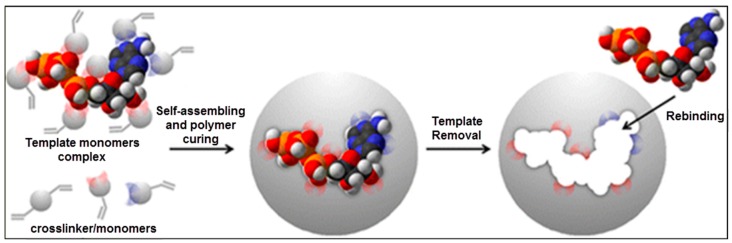
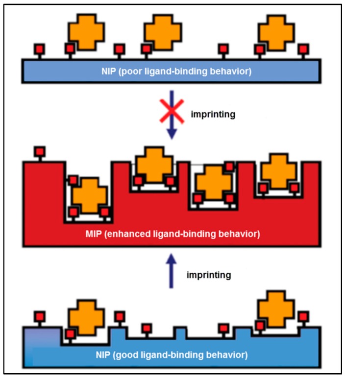
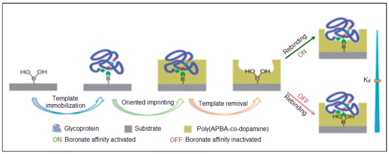
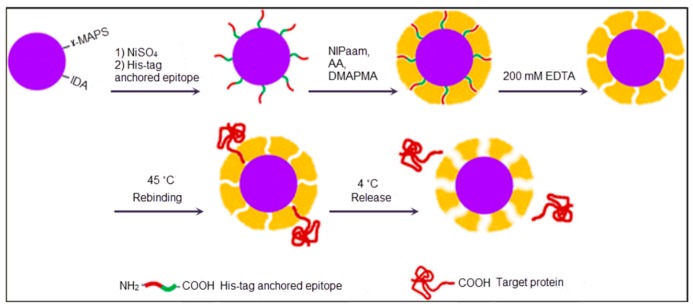
Molecular recognition has an important role in numerous living systems. One of the most important molecular recognition methods is molecular imprinting, which allows host compounds to recognize and detect several molecules rapidly, sensitively and selectively. Compared to natural systems, molecular imprinting methods have some important features such as low cost, robustness, high recognition ability and long term durability which allows molecularly imprinted polymers to be used in various biotechnological applications, such as chromatography, drug delivery, nanotechnology, and sensor technology. Sensors are important tools because of their ability to figure out a potentially large number of analytical difficulties in various areas with different macromolecular targets. Proteins, enzymes, nucleic acids, antibodies, viruses and cells are defined as macromolecules that have wide range of functions are very important. Thus, macromolecules detection has gained great attention in concerning the improvement in most of the studies. The applications of macromolecule imprinted sensors will have a spacious exploration according to the low cost, high specificity and stability. In this review, macromolecules for molecularly imprinted sensor applications are structured according to the definition of molecular imprinting methods, developments in macromolecular imprinting methods, macromolecular imprinted sensors, and conclusions and future perspectives. This chapter follows the latter strategies and focuses on the applications of macromolecular imprinted sensors. This allows discussion on how sensor strategy is brought to solve the macromolecules imprinting.

Keywords: macromolecule; molecular imprinting; sensor.


Conflict of interest statement

The authors declare no conflict of interest.

Figures

Figure 1
Figure 1
A schematic representation of the molecular imprinting method [14].
Figure 2
Figure 2
A schematic illustration of the alternative imprinting hypothesis [17].
Figure 3
Figure 3
A schematic illustration of the surface imprinting of glycoproteins [47].
Figure 4
Figure 4
A schematic representation of the epitope imprinting method [76].
Figure 5
Figure 5
The detection of lysozyme with lysozyme imprinted SPR sensor: (A) concentration dependence of lysozyme imprinted SPR sensor, (B) concentration versus SPR sensor response, (C) linear regions [89].
Figure 6
Figure 6
The (A) concentration dependency and (B) selectivity experiments of SPR sensor [90].
Figure 7
Figure 7
Schematic illustration of the preparation of lysozyme-imprinted polymer: (A) ({[2-(2-methacrylamido)ethyldithio]ethylcarbamoyl}methoxy)acetic acid structure, (B) protein-imprinted polymer preparation, (C) binding cavity created by the disulfide linkage reduction and (D) fluorophore introduction by the disulfide linkage reformation [91].
Figure 8
Figure 8
Atomic force microscopy pictures of (A) non-modified, (B) allyl mercaptan-modified, (C) hepatitis B surface antibody-imprinted SPR sensor [93].
Figure 9
Figure 9
Sensorgrams for the interaction between hepatitis B surface antibody positive human serum and hepatitis B surface antibody imprinted SPR sensor (A) reflectivity and (B) ΔR vs. time. [93].
Figure 10
Figure 10
(A) Glass slides preparation, (B) surface modification of SPR sensor, (C) micro-contact imprinting of prostate specific antigen, (D) surface modification of glass slides, (E) amino groups activation on glass slides and (F) prostate specific antigen immobilization onto the glass slides [94].
Figure 11
Figure 11
The (A) detection of total PSA antibody-modified gold nanoparticles after the PSA injection, (B) 4.69, 1.17, 0.29, 0 ng/mL on total PSA antibody immobilized surface and 150 ng/mL on IgG-immobilized surface, (C, linear, D, log scales) the calibration curves that came by from the assay [95].
Figure 12
Figure 12
The selectivity SPR sensor (AF): (i) Equilibrium by phosphate buffer, (ii) the analyte solutions application, and (iii) desorption with phosphate buffer that has 1 M NaCl [65].
Figure 13
Figure 13
The schematic model of solid phase imprinting to produce artificial antibodies [96].
Figure 14
Figure 14
The characterization of the glass and SPR sensor: The atomic force microscopy images of (A) myoglobin immobilized glass, (B) bare SPR sensor, (C) myoglobin imprinted SPR sensor [60].
Figure 15
Figure 15
The scheme and recovery rates of the QCM sensor [101].
Figure 16
Figure 16
Schematic representation of the protein C-imprinted QCM sensor [102].
Figure 17
Figure 17
Scheme of the formation of guanosine-imprints on SPR sensor surface [109].
Figure 18
Figure 18
The schematic representation of the cell-imprinted polydimethylsiloxane process: (A) The template preparation, (B) polymer imprinting with the template and (C) cell sorting with the microfluidic chip [115].
Figure 19
Figure 19
The scheme of the micro-contact imprinting of E. coli onto the polymer modified surfaces. The preparation of electrode surface (A), bacteria stamps (B), production of the micro-contact imprinting (C) [120].
Figure 20
Figure 20
A schematic model of the imprinted SPR and QCM sensors [121].
Figure 21
Figure 21
The surface morphologies of E. coli-imprinted (AC) SPR and (DF) QCM sensors [121].
Figure 22
Figure 22
The fundamental of the synthesis by using a solid-phase method [126].
Figure 23
Figure 23
The SPR sensor for virus detection [126].

References

    1. Wulff G., Sarhan A. Use of polymers with enzyme analogous structures for the resolution of racemates. Angew. Chem. Int. Ed. 1972;11:341–344.
    1. Wulff G., Gross T., Schonfeld R. Enzyme models based on molecularly imprinted polymers with strong esterase activity. Angew. Chem. Int. Ed. 1997;36:1962–1964. doi: 10.1002/anie.199719621. - DOI
    1. Algieri C., Drioli E., Guzzo L., Donato L. Bio-mimetic sensors based on molecularly imprinted membranes. Sensors. 2014;14:13863–13912. doi: 10.3390/s140813863. - DOI - PMC - PubMed
    1. Verma A., Nakade H., Simard J.M., Rotello V.M. Recognition and stabilization of peptide α-helices using templatable nanoparticle receptors. J. Am. Chem. Soc. 2004;126:10806–108074. doi: 10.1021/ja047719h. - DOI - PubMed
    1. Aubin-Tam M.-E., Hamad-Schifferli K. Gold nanoparticle-cytochrome c complexes: The effect of nanoparticle ligand charge on protein structure. Langmuir. 2005;21:12080–12084. doi: 10.1021/la052102e. - DOI - PubMed