

cGAS-STING-TBK1-IRF3/7 induced interferon-β contributes to the clearing of non tuberculous mycobacterial infection in mice
- PMID: 28422568
- PMCID: PMC5711412
- DOI: 10.1080/21505594.2017.1321191
cGAS-STING-TBK1-IRF3/7 induced interferon-β contributes to the clearing of non tuberculous mycobacterial infection in mice
Abstract
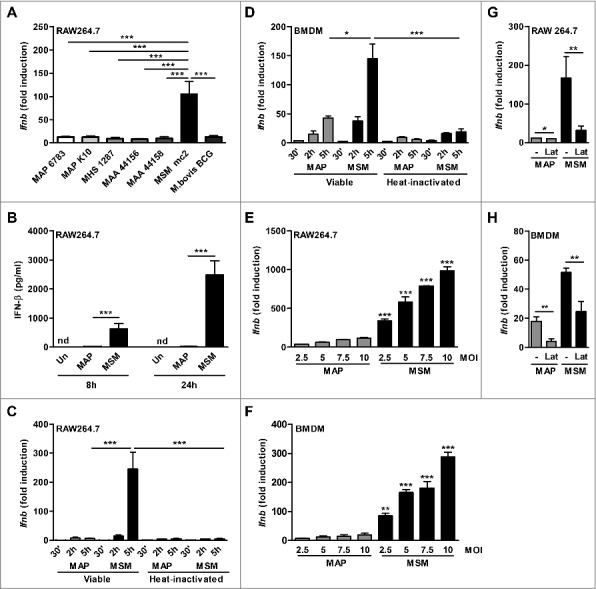
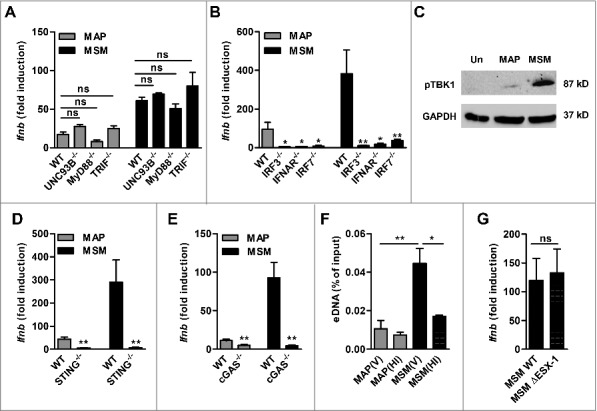
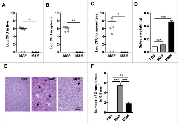
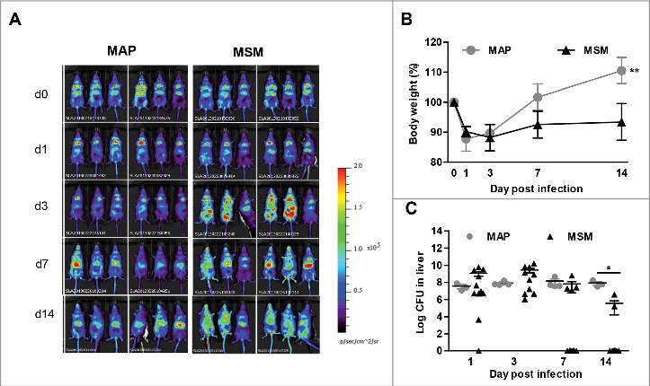
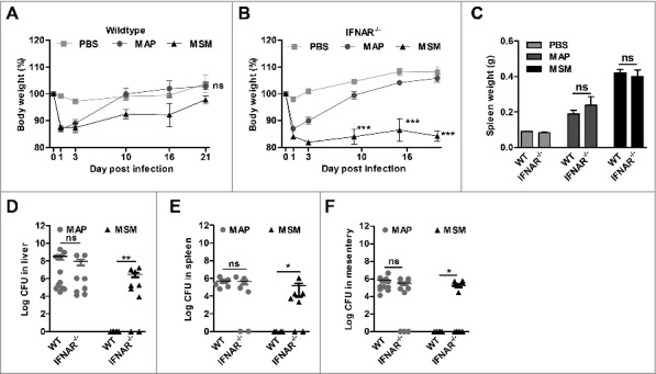
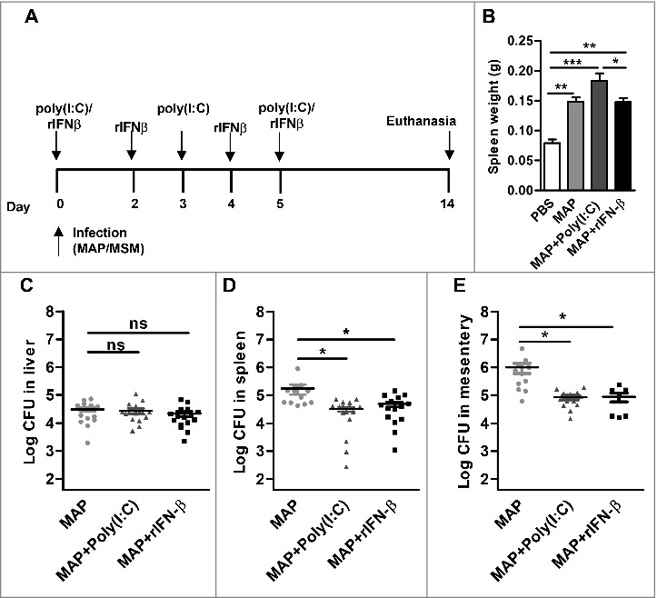
Type I interferons (IFN-I), such as IFN-α and IFN-β are important messengers in the host response against bacterial infections. Knowledge about the role of IFN-I in infections by nontuberculous mycobacteria (NTM) is limited. Here we show that macrophages infected with pathogens of the Mycobacterium avium complex produced significantly lower amounts of IFN-β than macrophages infected with the opportunistic pathogen M. smegmatis. To dissect the molecular mechanisms of this phenomenon, we focused on the obligate pathogen Mycobacterium avium ssp paratuberculosis (MAP) and the opportunistic M. smegmatis. Viability of both bacteria was required for induction of IFN-β in macrophages. Both bacteria induced IFN-β via the cGAS-STING-TBK1-IRF3/7-pathway of IFN-β activation. Stronger phosphorylation of TBK1 and higher amounts of extracellular bacterial DNA in the macrophage cytosol were found in M. smegmatis infected macrophages than in MAP infected macrophages. After intraperitoneal infection of mice, a strong Ifnb induction by M. smegmatis correlated with clearance of the bacteria. In contrast, MAP only induced weak Ifnb expression which correlated with bacterial persistence and increased number of granulomas in the liver. In mice lacking the type I interferon receptor we observed improved survival of M. smegmatis while survival of MAP was similar to that in wildtype mice. On the other hand, treatment of MAP infected wildtype mice with the IFN-I inducer poly(I:C) or recombinant IFN-β impaired the survival of MAP. This indicates an essential role of IFN-I in clearing infections by MAP and M. smegmatis. The expression level of IFN-I is decisive for transient versus persistent NTM infection.
Keywords: STING; Type I interferon; cGAS; extracellular DNA; mycobacterium paratuberculosis; mycobacterium smegmatis; non tuberculous mycobacteria.
Figures
Comment in
-
Interferon-β controls non-tuberculous mycobacterial infection in mice.Virulence. 2017 Oct 3;8(7):1085-1087. doi: 10.1080/21505594.2017.1341035. Epub 2017 Jun 12. Virulence. 2017. PMID: 28605283 Free PMC article. No abstract available.
Similar articles
-
African Swine Fever Virus Armenia/07 Virulent Strain Controls Interferon Beta Production through the cGAS-STING Pathway.J Virol. 2019 May 29;93(12):e02298-18. doi: 10.1128/JVI.02298-18. Print 2019 Jun 15. J Virol. 2019. PMID: 30918080 Free PMC article.
-
Modified vaccinia virus Ankara triggers type I IFN production in murine conventional dendritic cells via a cGAS/STING-mediated cytosolic DNA-sensing pathway.PLoS Pathog. 2014 Apr 17;10(4):e1003989. doi: 10.1371/journal.ppat.1003989. eCollection 2014 Apr. PLoS Pathog. 2014. PMID: 24743339 Free PMC article.
-
Nontypeable Haemophilus influenzae DNA stimulates type I interferon expression via STING signaling pathway.Biochim Biophys Acta Mol Cell Res. 2018 Apr;1865(4):665-673. doi: 10.1016/j.bbamcr.2018.01.011. Epub 2018 Feb 5. Biochim Biophys Acta Mol Cell Res. 2018. PMID: 29421524
-
The molecular mechanism of dsDNA sensing through the cGAS-STING pathway.Adv Immunol. 2024;162:1-21. doi: 10.1016/bs.ai.2024.02.003. Epub 2024 Mar 2. Adv Immunol. 2024. PMID: 38866436 Review.
-
The mechanism of double-stranded DNA sensing through the cGAS-STING pathway.Cytokine Growth Factor Rev. 2014 Dec;25(6):641-8. doi: 10.1016/j.cytogfr.2014.06.006. Epub 2014 Jun 22. Cytokine Growth Factor Rev. 2014. PMID: 25007740 Free PMC article. Review.
Cited by
-
Mycobacterium tuberculosis Infection of Retinal Endothelial Cells Induces Interferon Signaling Activation: Insights Into Tubercular Retinal Vasculitis.Invest Ophthalmol Vis Sci. 2025 Jul 1;66(9):48. doi: 10.1167/iovs.66.9.48. Invest Ophthalmol Vis Sci. 2025. PMID: 40668059 Free PMC article.
-
Myocardial Mitochondrial DNA Drives Macrophage Inflammatory Response through STING Signaling in Coxsackievirus B3-Induced Viral Myocarditis.Cells. 2023 Oct 31;12(21):2555. doi: 10.3390/cells12212555. Cells. 2023. PMID: 37947632 Free PMC article.
-
Endogenous Retroviruses as Modulators of Innate Immunity.Pathogens. 2023 Jan 19;12(2):162. doi: 10.3390/pathogens12020162. Pathogens. 2023. PMID: 36839434 Free PMC article. Review.
-
Activated cGAS/STING signaling elicits endothelial cell senescence in early diabetic retinopathy.JCI Insight. 2023 Jun 22;8(12):e168945. doi: 10.1172/jci.insight.168945. JCI Insight. 2023. PMID: 37345657 Free PMC article.
-
STING dependent BAX-IRF3 signaling results in apoptosis during late-stage Coxiella burnetii infection.Cell Death Dis. 2024 Mar 8;15(3):195. doi: 10.1038/s41419-024-06573-1. Cell Death Dis. 2024. PMID: 38459007 Free PMC article.
References
-
- Borden EC, Sen GC, Uze G, Silverman RH, Ransohoff RM, Foster GR, Stark GR. Interferons at age 50: past, current and future impact on biomedicine. Nat Rev Drug Discov 2007; 6:975-90; https://doi.org/10.1038/nrd2422 - DOI - PMC - PubMed
-
- Trinchieri G. Type I interferon: friend or foe? J Exp Med 2010; 207:2053-63; PMID:20837696; https://doi.org/10.1084/jem.20101664 - DOI - PMC - PubMed
-
- Boxx GM, Cheng G. The roles of type I interferon in bacterial infection. Cell Host Microbe 2016; 19:760-9; PMID:27281568; https://doi.org/10.1016/j.chom.2016.05.016 - DOI - PMC - PubMed
-
- Solodova E, Jablonska J, Weiss S, Lienenklaus S. Production of IFN-beta during listeria monocytogenes infection is restricted to monocyte/macrophage lineage. Plos One 2011; 6:e18543; https://doi.org/10.1371/journal.pone.0018543 - DOI - PMC - PubMed
-
- Stockinger S, Kastner R, Kernbauer E, Pilz A, Westermayer S, Reutterer B, Soulat D, Stengl G, Vogl C, Frenz T, et al. . Characterization of the Interferon-Producing Cell in Mice Infected with Listeria monocytogenes. Plos Pathogens 2009; 5:e1000355; PMID:19325882; https://doi.org/10.1371/journal.ppat.1000355 - DOI - PMC - PubMed
MeSH terms
Substances
LinkOut - more resources
Full Text Sources
Other Literature Sources
Medical
Molecular Biology Databases
Research Materials
Miscellaneous