. 2017 May 16;8(20):33444-33456.
doi: 10.18632/oncotarget.16800.

Fbxw7 haploinsufficiency loses its protection against DNA damage and accelerates MNU-induced gastric carcinogenesis

Affiliations

Fbxw7 haploinsufficiency loses its protection against DNA damage and accelerates MNU-induced gastric carcinogenesis

Yannan Jiang et al. Oncotarget. .

Abstract

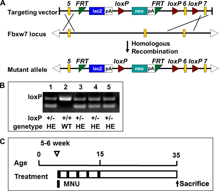
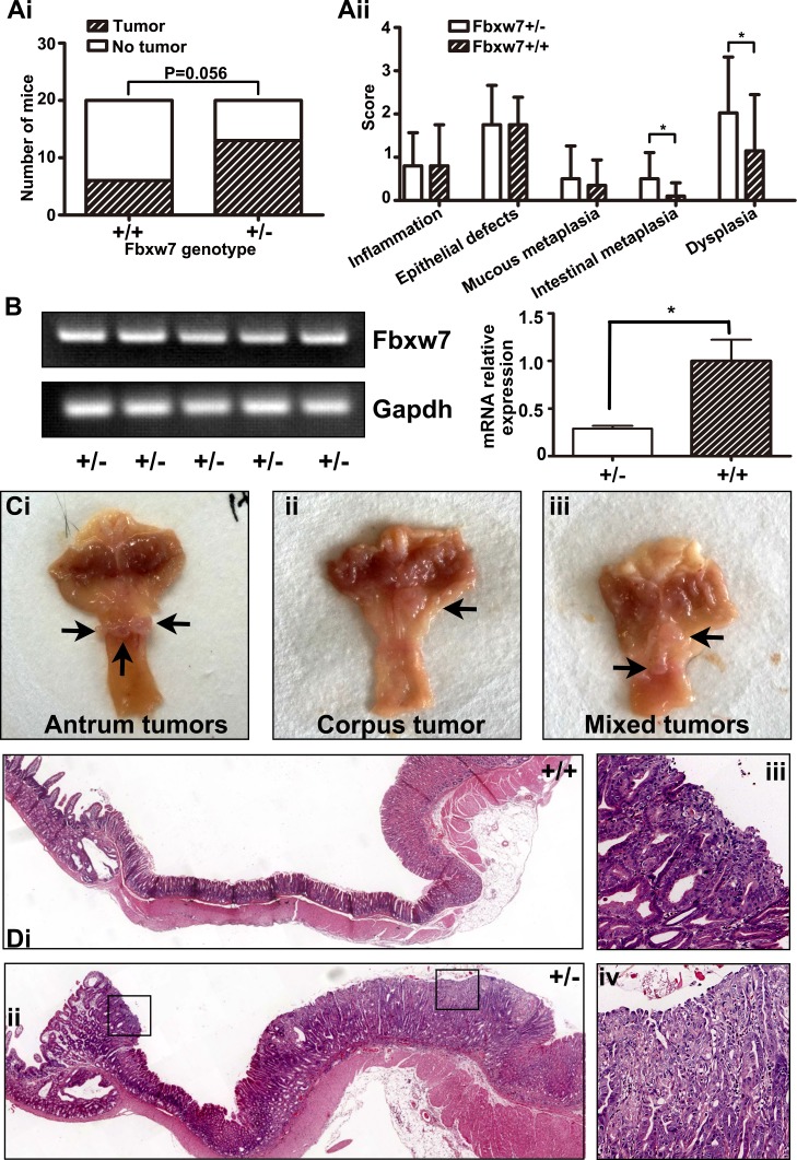
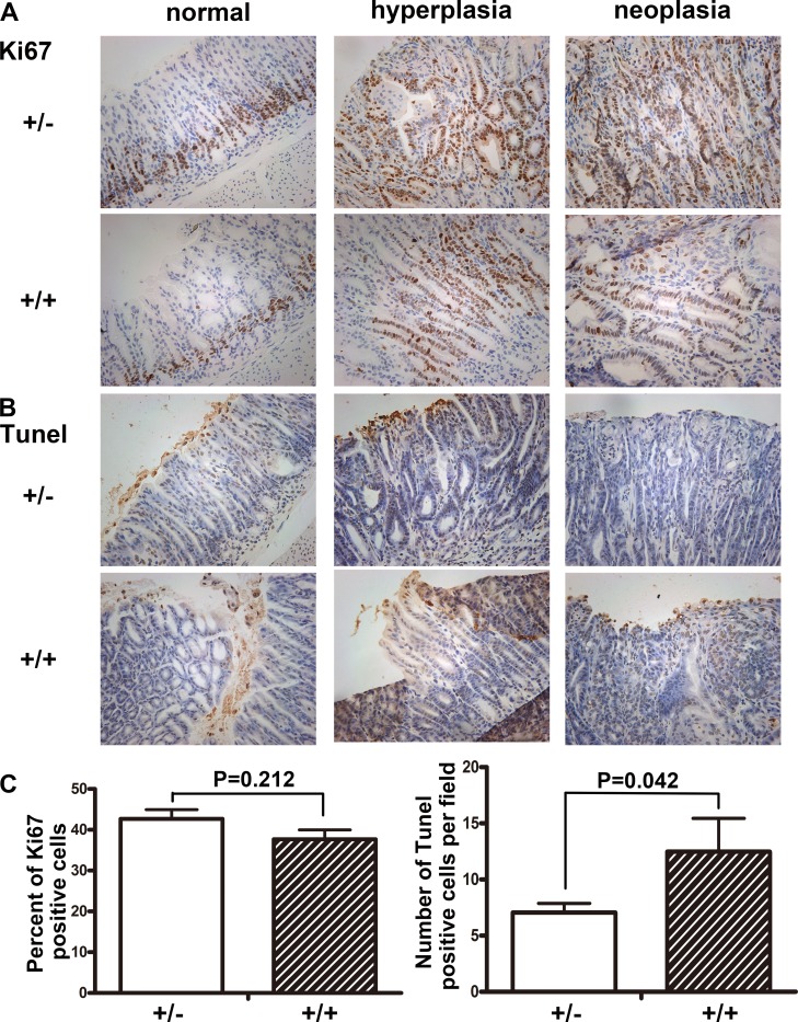
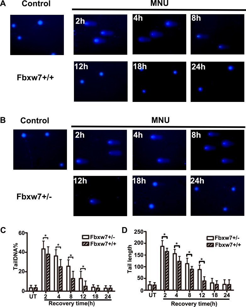
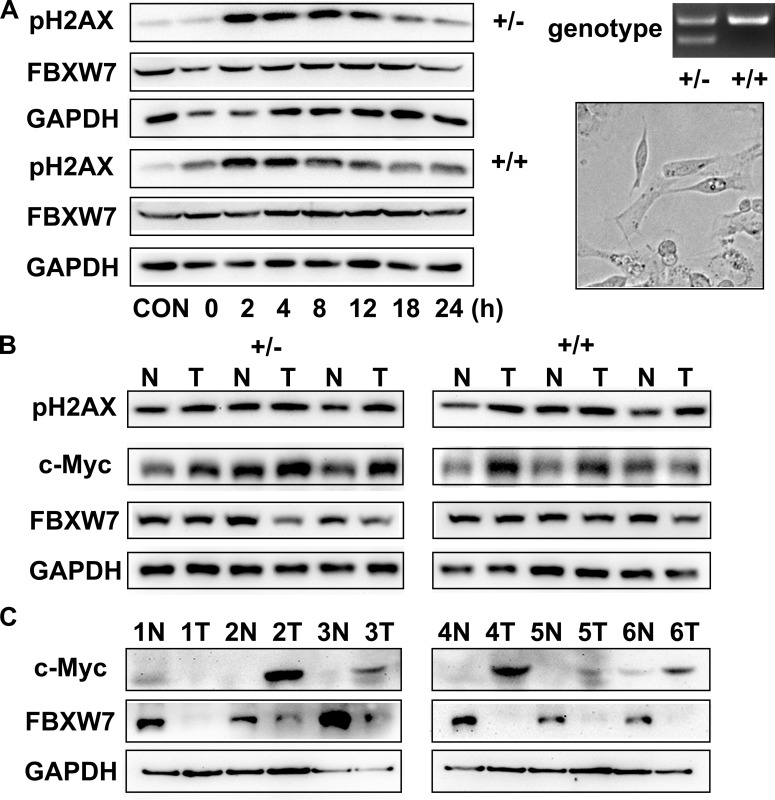
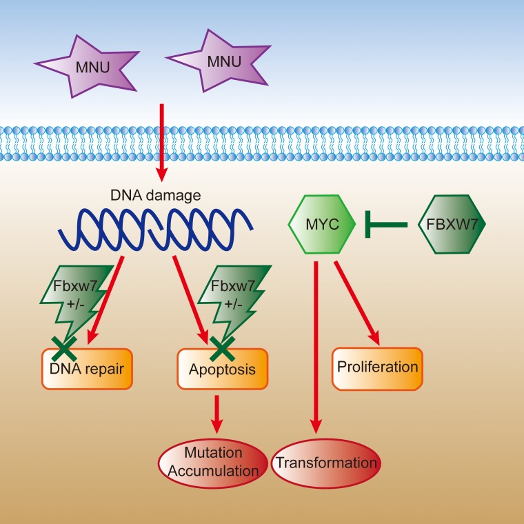
Fbxw7, a subunit of the SCF E3 ubiquitin ligase, recognizes oncoprotein substrates and leads to their proteasomal degradation. Fbxw7 acts as a tumor suppressor via inducing apoptosis and growth arrest in various kinds of tumors. To clarify the initiating role in gastric carcinogenesis as well as the histologic characterization of tumor in Fbxw7 allele haploinsufficient mice, we generated Fbxw7 heterozygous knockout mice (Fbxw7+/-) and treated them with chemical carcinogen N-methyl-N-nitrosourea (MNU) at 5-6 weeks of age. We also treated mouse embryo fibroblasts (MEFs) from Fbxw7+/- and Fbxw7+/+ mice with MNU and examined cell DNA damage via comet assay. The protein expression of Fbxw7 and its substrate c-Myc from mouse tumors, as well as human tumors sampled from six patients, were detected by Western blot. As results, the tumor incidence was obviously higher in Fbxw7+/- mice (13/20) than in Fbxw7+/+ mice (6/20) after 35-week observation. Intestinal metaplasia (P = 0.013) and dysplasia (P = 0.036) were more severe in Fbxw7+/- mice than in Fbxw7+/+ mice. The repair potential of DNA damage was suppressed in MEFs from Fbxw7+/- mice after MNU exposure. Increased c-Myc expression was accompanied by decreased Fbxw7 protein expression in tumor tissues from mouse and patients. In conclusion, Fbxw7 haploinsufficiency increased the risk of gastric carcinogenesis induced by MNU, which is associated with the accumulation of DNA damage as well as c-Myc oncoprotein.

Keywords: DNA damage; Fbxw7; N-Methyl-N-nitrosourea; gastric cancer; knockout mouse.


Conflict of interest statement

CONFLICTS OF INTEREST

None.

Figures

Figure 1
Figure 1. Fbxw7 knockout mice construction and treatment with MNU
(A) Schematic diagram of the creating Fbxw7 heterozygous knockout mice. (B) Genotyping using mouse tail DNA at the age of 2 weeks. PCR screening for the mutant allele revealed a band of 232bp for the wild type allele and 359bp for the mutant allele. (C) Experimental design of MNU treatment. The 5–6 weeks old mice were given MNU 240 mg/L in drinking water, every other week for a total exposure of 5 weeks.
Figure 2
Figure 2. Induction of gastric tumor development in the antrum in Fbxw7+/− mice
(A) Incidence (Ai) and pathological score (Aii) of gastric tumor formation in Fbxw7+/− and Fbxw7+/+ mice at the age of 35 weeks with MNU-based treatment (n = 20 each genotype). (B) mRNA expression of Fbxw7 from tumors of Fbxw7+/− mice (n = 5 each genotype, left figure is from the semi-quantitative examination, and right figure is from the real-time RT-PCR assay). (C) Gross morphology of visible tumors (black arrows) in the gastric antrum (Ci), corpus (Cii), or both (“mixed”, Ciii) in Fbxw7+/− mice enlarged. (D) Representative photographs of macroscopic views of the entire gastric mucosa in Fbxw7+/+ (Di) and Fbxw7+/− (Dii) mice and two microscopic view of an gastric tumor in the Fbxw7+/− mouse (Diii, Div). The 2-tailed X2 test was used to determine the significance of the tumor incidence. The non-parametric test was used to determine the significance of the difference between pathological scores of each group. “*” refers to statistically significant (P < 0.05).
Figure 3
Figure 3. Immunohistochemistry staining of Ki67 and TUNEL in antrum
Sections of antral tissues were prepared from Fbxw7+/− and Fbxw7+/+ mice. From left to right were: normal, hyperplastic and neoplastic tissues. Cell proliferation was determined by Ki67 staining. Cell apoptosis was determined by TUNEL. The X2 or T test was used to determine the significance of Ki67 and TUNEL studies.
Figure 4
Figure 4. Bimodal pattern of H2AX phosphorylation after treatment with MNU
(A, B) pH2AX foci in MEFs from Fbxw7+/+ (A) and Fbxw7+/− (B) after MNU treatment. The cells were collected and fixed at the indicated time points after treatment. (C, D) The dynamics of pH2AX foci formation after treatment as shown in (A) and (B). Data are presented as mean ± SD, from three independent experiments. The X2 or T test was used to determine the significance of pH2AX positive incidence and intensity. “*” refers to statistically significant (P < 0.05).
Figure 5
Figure 5. Detection of DNA damage by comet assay
(A, B) The comet images of MEFs from Fbxw7+/+ (A) and Fbxw7+/− (B) at given time after MNU treatment. (C, D) The repair kinetics of DNA damage, which were expressed as the comets tailDNA% (C) and tail length (D). The data are the means and standard deviation from three experiments, and more than 50 comets were analyzed in each experiment. The T test was used to determine the significance of comet assay. “*” refers to statistically significant (P < 0.05).
Figure 6
Figure 6. Loss of Fbxw7 expression and overexpression of c-Myc in gastric cancer
(A) Western blot for pH2AX expression at indicated time points after MNU treatment (left). Genotyping and microscopic view of Fbxw7+/+ and Fbxw7+/− MEFs (right). (B, C) Western blot analyses were performed using total protein lysates extracted from antral mucosa from mice (B) and from patients (C).
Figure 7
Figure 7. The schematic figure of somatic cells transformation induced by MNU in Fbxw7 haploinsufficiency condition
Cell DNA is damaged under the MNU exposure. Due to the haploinsufficiency of Fbxw7, it is difficult to repair mutated DNA as well as proceed apoptosis. Meanwhile, insufficient FBXW7 level results in elevated downstream c-MYC expression and promoted cell transformation and proliferation.

Similar articles

Cited by

References

    1. Ferlay J, Soerjomataram I, Dikshit R, Eser S, Mathers C, Rebelo M, Parkin DM, Forman D, Bray F. Cancer incidence and mortality worldwide: Sources, methods and major patterns in GLOBOCAN 2012. International journal of cancer Journal international du cancer. 2015;136:E359–386. - PubMed
    1. Wang Z, Inuzuka H, Zhong J, Wan L, Fukushima H, Sarkar FH, Wei W. Tumor suppressor functions of FBW7 in cancer development and progression. FEBS letters. 2012;586:1409–1418. - PMC - PubMed
    1. Tan Y, Sangfelt O, Spruck C. The Fbxw7/hCdc4 tumor suppressor in human cancer. Cancer letters. 2008;271:1–12. - PubMed
    1. Welcker M, Clurman BE. FBW7 ubiquitin ligase: a tumour suppressor at the crossroads of cell division, growth and differentiation. Nature reviews Cancer. 2008;8:83–93. - PubMed
    1. Cheng Y, Li G. Role of the ubiquitin ligase Fbw7 in cancer progression. Cancer metastasis reviews. 2012;31:75–87. - PubMed

MeSH terms

Substances