. 2017 May 30;8(22):36383-36398.
doi: 10.18632/oncotarget.16756.

Calmodulin promotes matrix metalloproteinase 9 production and cell migration by inhibiting the ubiquitination and degradation of TBC1D3 oncoprotein in human breast cancer cells

Affiliations

Calmodulin promotes matrix metalloproteinase 9 production and cell migration by inhibiting the ubiquitination and degradation of TBC1D3 oncoprotein in human breast cancer cells

Huzi Zhao et al. Oncotarget. .

Abstract

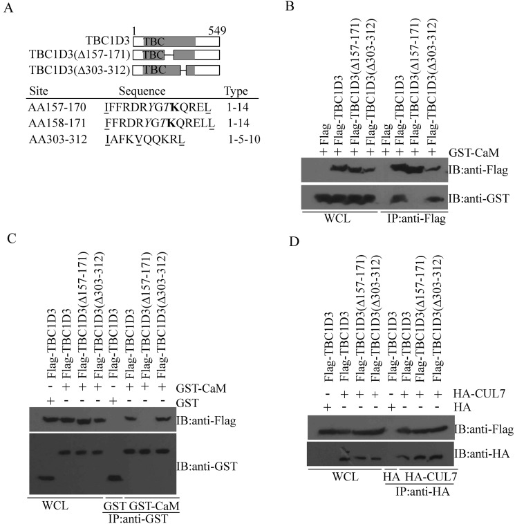
The hominoid oncoprotein TBC1D3 enhances growth factor (GF) signaling and GF signaling, conversely, induces the ubiquitination and subsequent degradation of TBC1D3. However, little is known regarding the regulation of this degradation, and the role of TBC1D3 in the progression of tumors has also not been defined. In the present study, we demonstrated that calmodulin (CaM), a ubiquitous cellular calcium sensor, specifically interacted with TBC1D3 in a Ca2+-dependent manner and inhibited GF signaling-induced ubiquitination and degradation of the oncoprotein in both cytoplasm and nucleus of human breast cancer cells. The CaM-interacting site of TBC1D3 was mapped to amino acids 157~171, which comprises two 1-14 hydrophobic motifs and one lysine residue (K166). Deletion of these motifs was shown to abolish interaction between TBC1D3 and CaM. Surprisingly, this deletion mutation caused inability of GF signaling to induce the ubiquitination and subsequent degradation of TBC1D3. In agreement with this, we identified lysine residue 166 within the CaM-interacting motifs of TBC1D3 as the actual site for the GF signaling-induced ubiquitination using mutational analysis. Point mutation of this lysine residue exhibited the same effect on TBC1D3 as the deletion mutant, suggesting that CaM inhibits GF signaling-induced degradation of TBC1D3 by occluding its ubiquitination at K166. Notably, we found that TBC1D3 promoted the expression and activation of MMP-9 and the migration of MCF-7 cells. Furthermore, interaction with CaM considerably enhanced such effect of TBC1D3. Taken together, our work reveals a novel model by which CaM promotes cell migration through inhibiting the ubiquitination and degradation of TBC1D3.

Keywords: TBC1D3; calmodulin; cell migration; protein degradation; protein ubiquitination.


Conflict of interest statement

CONFLICTS OF INTEREST

The authors declare no conflicts of interest.

Figures

Figure 1
Figure 1. CaM inhibits the ubiquitination and degradation of TBC1D3 in both cytoplasm and the nucleus in response to FCS stimulation
(AB) MCF-7 and BT549 cells (left and right panels, respectively) were transfected with Flag-TBC1D3 alone (B) or together with GST-CaM or control GST vector (A). After 20 h, the cells were starved in serum-free medium with or without W7 (90 μM) for 3 h, and then stimulated by the addition of 10% fetal calf serum (FCS) in the presence of cycloheximide (25 μg/ml) for the indicated time. Cell extracts were resolved by SDS-PAGE and immunoblotted with anti-Flag (TBC1D3), anti-GST and anti-GAPDH antibodies. The number below each blot represents the percent TBC1D3 remaining at the indicated time point of FCS treatment normalized to the zero time point. (C) MCF-7 and BT549 cells (left and right panels, respectively) were transfected with Flag-TBC1D3 alone or together with GST-CaM. After 20 h, the cells were serum-starved in the presence of MG132 for 6 h, and then stimulated with (+) or without (−) 10% FCS for 20 min. Lysates were immunoprecipitated (IP) with anti-Flag antibody. After SDS-PAGE, Flag-TBC1D3, GST-CaM and polyubiquitinated TBC1D3 were immunoblotted (IB) with anti-Flag, anti-GST and anti-ubiquitin (Ubi) antibodies, respectively. Molecular weight markers are shown on right in kD. WCL, whole cell lysate. (D) MCF-7 and BT549 cells (left and right panels, respectively) were transfected with Flag-TBC1D3 alone or together with GST-CaM. After 20 h, the cells were starved in serum-free medium for 3 h, stimulated with (+) or without (−) 10% fetal calf serum (FCS) in the presence of cycloheximide (25 μg/ml) for 3 h, and then subjected to cell fractionation. Cytoplasmic and nucleic extracts were resolved by SDS-PAGE and immunoblotted with anti-Flag, anti-GST, anti-PARP-1 and anti-GAPDH antibodies.
Figure 2
Figure 2. CaM associates with TBC1D3 in a Ca2+-dependent manner
MCF-7 (A, B and D) and BT549 (C) cells were co-transfected with Flag-TBC1D3 together with GST-CaM or control GST vector (A and C), or with GST-CaM together with Flag-TBC1D3 or control Flag vector (B and D), and then treated with or without EGTA (2 mM). Lysates were immunoprecipitated (IP) with anti-GST (A and C) or anti-Flag (B and D) antibodies. After SDS-PAGE, Flag-TBC1D3 and GST-CaM/GST were immunoblotted (IB) with anti-Flag and anti-GST antibodies, respectively. Molecular weight markers are shown on right in kD. WCL, whole cell lysate.
Figure 3
Figure 3. Mapping of the CaM-interacting site in TBC1D3
(A) Schematic representation of full-length TBC1D3 and various deletion mutants in top panel. The length or internal deletion (Δ) of each isoform in amino acids is indicated. TBC/Rab GAP homology domain is shown in gray. Three potential CaM-interacting motifs, two potential phosphorylatable amino acid residues and one potential ubiquitination site in TBC1D3 were identified in bottom panel. The position (in amino acids (AA)), sequence of the CaM-interacting sites (with conserved hydrophobic residues underlined, phosphorylatable residues italicized, and ubiquitination site boldfaced), and motif type are shown for each site. (BD) MCF-7 cells were co-transfected with GST-CaM (B), GST-CaM/control GST (C), or HA-CUL7/control HA (D), together with control Flag vector, Flag-TBC1D3, or TBC1D3 mutants harboring internal deletions of the indicated residues. Lysates were immunoprecipitated (IP) with anti-Flag (B), anti-GST (C), or anti-HA (D) antibodies. After SDS-PAGE, Flag-TBC1D3, GST-CaM/GST, and HA-CUL7 were immunoblotted (IB) with anti-Flag, anti-GST and anti-HA antibodies, respectively. WCL, whole cell lysate.
Figure 4
Figure 4. Mutation of the ubiquitination site K166 abolishes the ubiquitination and degradation of TBC1D3 in response to FCS stimulation
(AD) MCF-7 cells were transfected with Flag-tagged wild-type (A, B), internal deletion mutant (A, B), or point mutants (C, D) of TBC1D3. After 20 h, the cells were starved in serum-free medium for 3 h, and then stimulated with or without 10% fetal calf serum (FCS) in the presence of cycloheximide (25 μg/ml) for the indicated times. Cell extracts were resolved by SDS-PAGE and immunoblotted with anti-Flag and anti-GAPDH antibodies. The bar graphs B and D are derived from densitometric analysis of Western blots as typified in A and C, respectively. Expression of TBC1D3 and its mutant is normalized to GAPDH. The initial level of TBC1D3 expression in each group is set to 100%. The data are presented as means ± SD of three independent experiments (*p < 0.05). (E) MCF-7 cells were transfected with Flag-tagged wild-type or indicated point mutants of TBC1D3. After 20 h, the cells were serum-starved in the presence of MG132 for 6 h, and then stimulated with (+) or without (−) 10% FCS for 20 min. Lysates were immunoprecipitated (IP) with anti-Flag antibody. After SDS-PAGE, non- and poly-ubiquitinated forms of Flag-TBC1D3 and its point mutants were immunoblotted (IB) with anti-Flag and anti-ubiquitin (Ubi) antibodies, respectively. Molecular weight markers are shown on right in kD. (F) MCF-7 cells were co-transfected with GST-CaM together with control Flag vector, Flag-tagged wild-type or indicated point mutant of TBC1D3. Lysates were immunoprecipitated (IP) with anti-Flag antibody. After SDS-PAGE, Flag-tagged wild-type or indicated point mutants of TBC1D3 and GST-CaM were immunoblotted (IB) with anti-Flag and anti-GST antibodies, respectively. WCL, whole cell lysate.
Figure 5
Figure 5. TBC1D3 overexpression promotes the expression and activation of MMP-9 and the migration of MCF-7 cells
(A) MCF-7 cells were transfected with Flag-TBC1D3 or control Flag vector. After 20 h, the confluent monolayer of the transfected cells were scratched and then allowed to migrate into the wound area for the indicated times. Photographs were taken at a magnification of × 200. Scale bar, 50 μm. (B) The bar graph is derived from scratch wound assay as typified in A. The rate of cell migration was analyzed based on the percentage of the distance of cell migration into the wound area at the indicated time points over the initial wound width in each group, using ImageJ software. The data are presented as means ± SD of three independent experiments (*p < 0.05). (C) MCF-7 and BT549 cells were transfected with Flag-TBC1D3 or control Flag vector. After 20 h, the transfected cells were added into the upper chambers with non-coated membrane (8-μm and 12-μm pore size for MCF-7 and BT549 cells, respectively) and complete medium containing 20% FCS into the lower chambers of transwells. Following incubation for 24 h, cells migrating from the top side to the bottom side of the membrane were fixed, stained, and then photographed at a magnification of X 200. Scale bar, 50 μm. (D) The bar graph is derived from transwell cell migration assay as typified in C. Five randomly selected fields of cells were photographed and counted using ImageJ software. The data are presented as means ± SD of three independent experiments (*p < 0.05). (E) MCF-7 cells were transfected with Flag-TBC1D3 or control Flag vector, and then starved in serum-free medium for 24 h. Whole cell lysates were immunoblotted for MMP-9 (including pro-MMP-9 and active MMP-9), Flag-TBC1D3, and GAPDH as a loading control. MMP-9 activity in concentrated conditioned medium was detected by gelatin zymography. (F) MCF-7 cells were transfected with Flag-TBC1D3 or control Flag vector, and then starved in serum-free medium for 24 h. Total RNA was extracted from the transfected cells and reverse-transcribed to cDNA. Subsequently, expression of MMP-9 mRNA in each group was analyzed by real-time quantitative PCR and normalized to GAPDH. The data are presented as means ± SD of three independent experiments (*p < 0.05).
Figure 6
Figure 6. CaM enhances the TBC1D3-induced expression and activation of MMP-9 and migration of MCF-7 cells
(A) MCF-7 cells were transfected with the indicated plasmids. After 20 h, the transfected cells were added into the upper chambers with non-coated membrane (8-μm pore size) and complete medium containing 20% FCS into the lower chambers of transwells. Following incubation for 24 h, cells migrating from the top side to the bottom side of the membrane were fixed, stained, and then photographed at a magnification of X 200. Scale bar, 50 μm. (B) The bar graph is derived from transwell cell migration assay as typified in A. Five randomly selected fields of cells were photographed and counted using ImageJ software. The data are presented as means ± SD of three independent experiments (*p < 0.05). (C) MCF-7 cells were transfected with the indicated plasmids, and then starved in serum-free medium for 24 h. Whole cell extracts were immunoblotted for MMP-9 (including pro-MMP-9 and active MMP-9), Flag-TBC1D3, GST, GST-CaM and GAPDH. MMP-9 activity in concentrated conditioned medium was detected by gelatin zymography. (DF) The bar graphs D, E and F are derived from densitometric analysis of Western blots as typified in C. Expression Pro-MMP-9 (D) and active MMP-9 (E) as well as MMP-9 activity (F) is normalized to GAPDH. The data are presented as means ± SD of three independent experiments (*p < 0.05).

References

    1. Ciechanover A. Intracellular protein degradation: from a vague idea through the lysosome and the ubiquitin-proteasome system and onto human diseases and drug targeting. Bioorg Med Chem. 2013;21:3400–10. doi: 10.1016/j.bmc.2013.01.056. - DOI - PubMed
    1. Knecht E, Aguado C, Carcel J, Esteban I, Esteve JM, Ghislat G, Moruno JF, Vidal JM, Saez R. Intracellular protein degradation in mammalian cells: recent developments. Cell Mol Life Sci. 2009;66:2427–43. doi: 10.1007/s00018-009-0030-6. - DOI - PMC - PubMed
    1. Rubinsztein DC. The roles of intracellular protein-degradation pathways in neurodegeneration. Nature. 2006;443:780–6. doi: 10.1038/nature05291. - DOI - PubMed
    1. Ciechanover A. Proteolysis: from the lysosome to ubiquitin and the proteasome. Nat Rev Mol Cell Biol. 2005;6:79–87. doi: 10.1038/nrm1552. - DOI - PubMed
    1. Pickart CM, Eddins MJ. Ubiquitin: structures, functions, mechanisms. Biochim Biophys Acta. 2004;1695:55–72. doi: 10.1016/j.bbamcr.2004.09.019. - DOI - PubMed

MeSH terms