. 2017 Apr 20;2(8):e93136.
doi: 10.1172/jci.insight.93136.

Steroid metabolome analysis reveals prevalent glucocorticoid excess in primary aldosteronism

Affiliations

Steroid metabolome analysis reveals prevalent glucocorticoid excess in primary aldosteronism

Wiebke Arlt et al. JCI Insight. .

Abstract

Background: Adrenal aldosterone excess is the most common cause of secondary hypertension and is associated with increased cardiovascular morbidity. However, adverse metabolic risk in primary aldosteronism extends beyond hypertension, with increased rates of insulin resistance, type 2 diabetes, and osteoporosis, which cannot be easily explained by aldosterone excess.

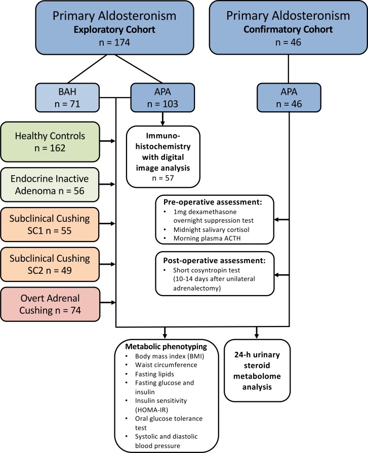
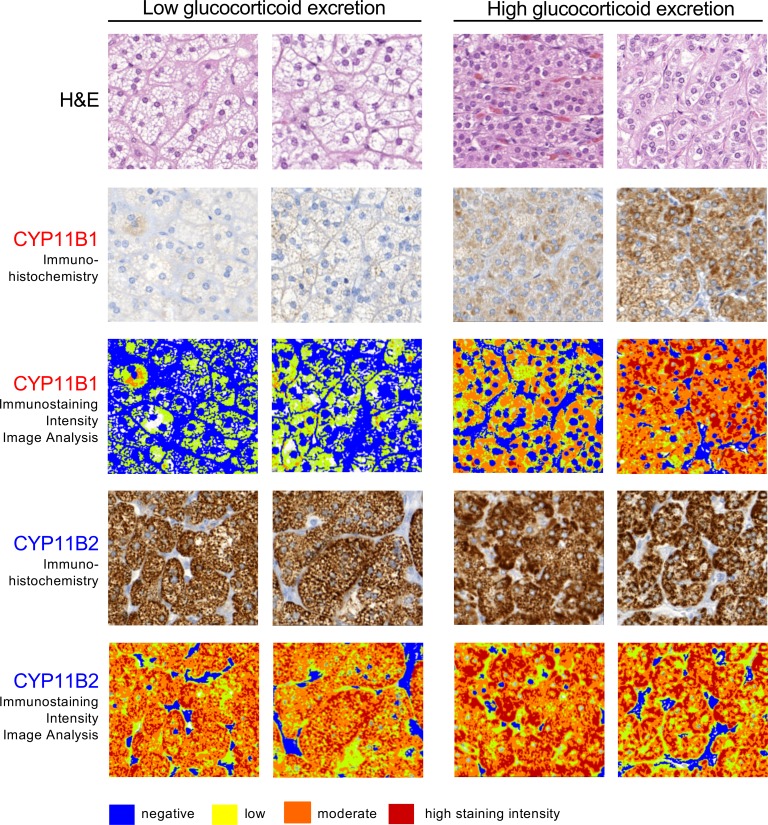
Methods: We performed mass spectrometry-based analysis of a 24-hour urine steroid metabolome in 174 newly diagnosed patients with primary aldosteronism (103 unilateral adenomas, 71 bilateral adrenal hyperplasias) in comparison to 162 healthy controls, 56 patients with endocrine inactive adrenal adenoma, 104 patients with mild subclinical, and 47 with clinically overt adrenal cortisol excess. We also analyzed the expression of cortisol-producing CYP11B1 and aldosterone-producing CYP11B2 enzymes in adenoma tissue from 57 patients with aldosterone-producing adenoma, employing immunohistochemistry with digital image analysis.

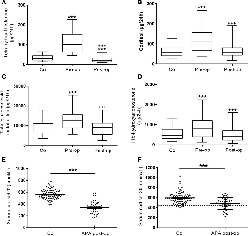
Results: Primary aldosteronism patients had significantly increased cortisol and total glucocorticoid metabolite excretion (all P < 0.001), only exceeded by glucocorticoid output in patients with clinically overt adrenal Cushing syndrome. Several surrogate parameters of metabolic risk correlated significantly with glucocorticoid but not mineralocorticoid output. Intratumoral CYP11B1 expression was significantly associated with the corresponding in vivo glucocorticoid excretion. Unilateral adrenalectomy resolved both mineralocorticoid and glucocorticoid excess. Postoperative evidence of adrenal insufficiency was found in 13 (29%) of 45 consecutively tested patients.

Conclusion: Our data indicate that glucocorticoid cosecretion is frequently found in primary aldosteronism and contributes to associated metabolic risk. Mineralocorticoid receptor antagonist therapy alone may not be sufficient to counteract adverse metabolic risk in medically treated patients with primary aldosteronism.

Funding: Medical Research Council UK, Wellcome Trust, European Commission.

Keywords: Cardiology; Endocrinology.


Conflict of interest statement

Conflict of interest: W. Arlt is the holder of a patent for the differential diagnosis of adrenal tumors (PCT/GB2010/000274); all other authors have no conflict of interest to declare.

Figures

Figure 1
Figure 1. CONSORT diagram visualizing the included patient and comparator groups and the performed diagnostic work-up.
After analysis of the results from the exploratory cohort, 46 patients were prospectively recruited with consecutive enrollment into the confirmatory cohort that underwent additional pre-and postoperative assessment of cortisol production. APA, unilateral aldosterone-producing adenoma; ACTH, adrenocorticotropic hormone; BAH, bilateral adrenal hyperplasia; HOMA-IR, homeostasis model assessment of insulin resistance.
Figure 2
Figure 2. Heatmap visualizations of steroid metabolome profiling results in 174 primary aldosteronism patients.
Steroid metabolite excretion in 24-hour urine was measured by gas chromatography-mass spectrometry in selected-ion-monitoring mode. Heatmap visualizations depict log-transformed steroid metabolite excretion values in the primary aldosteronism patients (group: black) in comparison to 162 healthy controls (group: white). (A) Mineralocorticoid and mineralocorticoid precursor metabolites ordered according to increasing amounts of tetrahydroaldosterone (THAldo) excretion. (B) Glucocorticoid metabolites in order of increasing amounts of cortisol excretion. Scale and color code were chosen separately for each panel according to the respective range of observed values. THA, tetrahydro-11-dehydrocorticosterone; THB, tetrahydrocorticosterone; THDOC, tetrahydro-11-deoxycorticosterone; THF, tetrahydrocortisol; THE, tetrahydrocortisone.
Figure 3
Figure 3. Steroid metabolite excretion in primary aldosteronism in comparison to healthy controls and patients with endocrine-inactive and cortisol-producing adrenal adenomas.
The panels show the 24-hour urinary excretion of tetrahydroaldosterone (A), cortisol (B), total glucocorticoid metabolites (C), and the major adrenal androgen metabolite 11β-hydroxyandrosterone (D) in primary aldosteronism patients (PA; n = 174) in comparison to healthy controls (Co; n = 162), patients with endocrine-inactive adrenal adenoma (EIA; n = 56), patients with subclinical Cushing’s (differentiated into 2 groups: SC1 (n = 55), morning cortisol after 1 mg dexamethasone overnight > 50 and < 138 nmol/l; SC2 (n = 49), morning cortisol >138 nmol/l), and overt adrenal Cushing’s syndrome patients (Cu; n = 47). Boxes represent median and interquartile range, whiskers represent 5th and 95th centiles. **P < 0.01 versus controls, ***P < 0.001 versus controls. Comparisons between groups were made with linear regression models to adjust for age and sex in comparisons between all 6 groups.
Figure 4
Figure 4. Steroid excretion in 46 patients with primary aldosteronism due to aldosterone-producing adenoma (APA) before and after unilateral adrenalectomy in comparison to healthy controls (n = 162).
The panels show 24-hour urinary excretion of tetrahydroaldosterone (A), cortisol (B), total glucocorticoid metabolites (C), and 11β-hydroxyandrosterone (D). ***P < 0.001 versus controls (Co); +++P < 0.001 versus preoperative. Boxes represent median and interquartile range, whiskers represent 5th and 95th centiles. Panels E and F show individual values and mean ± SEM for serum cortisol at baseline and 30 minutes after cosyntropin stimulation in 46 primary aldosteronism patients tested 2 weeks postoperatively, in comparison to 82 healthy controls. The dotted line represents the diagnostic cut-off for adrenal insufficiency (serum cortisol 30 minutes after 250 μg cosyntropin < 15.6 μg/dl [<430 nmol/l], equivalent to the 5th centile of the cortisol response in the 82 healthy controls). ***P < 0.001 versus controls. Comparisons of steroid excretion before and after unilateral adrenalectomy were made using 2-sided Wilcoxon’s signed-rank test. Linear models were also fitted comparing preoperative and postoperative log-transformed steroid-metabolite measures to controls, adjusting for age and sex.
Figure 5
Figure 5. Immunohistochemistry with digital image analysis for steroidogenic enzyme expression in aldosterone-producing adrenal adenoma tissue in relation to in vivo 24-hour glucocorticoid excretion.
A tissue microarray with adenoma tissue from 57 patients was studied by immunohistochemistry for expression of CYP11B1, required for cortisol and 11β-hydroxyandrostenedione synthesis, and CYP11B2, the enzyme responsible for aldosterone synthesis, followed by digital image analysis for quantification of staining intensity. Representative immunohistochemistry examples from 2 patients with in vivo glucocorticoid excretion within the lowest and highest quartiles, respectively, are shown (total original magnification in all panels, ×20).

References

    1. Funder JW, et al. The management of primary aldosteronism: Case detection, diagnosis, and treatment: an Endocrine Society Clinical Practice Guideline. J Clin Endocrinol Metab. 2016;101(5):1889–1916. doi: 10.1210/jc.2015-4061. - DOI - PubMed
    1. Hannemann A, et al. Screening for primary aldosteronism in hypertensive subjects: results from two German epidemiological studies. Eur J Endocrinol. 2012;167(1):7–15. doi: 10.1530/EJE-11-1013. - DOI - PubMed
    1. Hannemann A, Wallaschofski H. Prevalence of primary aldosteronism in patient’s cohorts and in population-based studies--a review of the current literature. Horm Metab Res. 2012;44(3):157–162. doi: 10.1055/s-0031-1295438. - DOI - PubMed
    1. Rossi GP, et al. A prospective study of the prevalence of primary aldosteronism in 1,125 hypertensive patients. J Am Coll Cardiol. 2006;48(11):2293–2300. doi: 10.1016/j.jacc.2006.07.059. - DOI - PubMed
    1. Choi M, et al. K+ channel mutations in adrenal aldosterone-producing adenomas and hereditary hypertension. Science. 2011;331(6018):768–772. doi: 10.1126/science.1198785. - DOI - PMC - PubMed