

Hypoxia-regulated human periodontal ligament cells via Wnt/β-catenin signaling pathway
- PMID: 28422843
- PMCID: PMC5406059
- DOI: 10.1097/MD.0000000000006562
Hypoxia-regulated human periodontal ligament cells via Wnt/β-catenin signaling pathway
Abstract
Background: The aim of this study is to investigate the effects of hypoxia on the proliferation, morphology, migration ability, hypoxia inducible factor (HIF) 1 (HIF-1) expression, and the relationship with Wnt/β-catenin signaling of human periodontal ligament cells (hPDLCs) in vitro.
Methods: hPDLCs (4th passage) cultured by the tissue culture method were randomly assigned to slight (5% O2), severe hypoxia (1% O2) groups, and the control (21% O2) group, respectively. From 1st to 7th day, the optical density values were detected, and the growth curve was described. Wound healing assay was done to observe the migration ability of hPDLCs under various O2 conditions. Then reverse transcription quantitative real-time polymerase chain reaction (RT-qPCR) was used to detect the expression of cementum-related genes and Wnt signaling pathway-related genes. Further, RT-qPCR, Western blot, and immunofluorescence staining method were constructed to show HIF expressions under different O2 concentrations in hPDLCs.
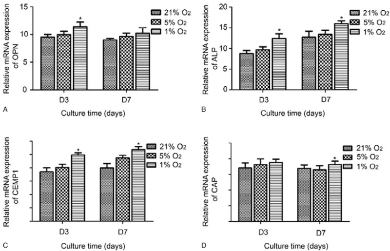
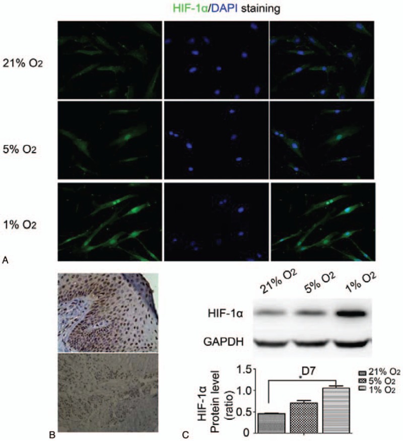
Results: The growth rate of hPDLCs decreased with the reduction of O2 content by degree, and the morphology of hPDLCs changed in different O2 contents. Besides, hPDLCs migrate faster in 21% and 5% O2 than in 1% O2, and both the expressions of cementum-related genes and Wnt signaling pathway-related genes were raised under hypoxic conditions. In addition, with the reduction of O2 concentration, the messenger RNA and protein level expression of HIF were all increased, and HIF was gradually transported from cytoplasm into the nucleus and in 1% O2 concentration, it was mainly expressed in the nucleus.
Conclusion: This finding demonstrated that hypoxia was capable of suppressing the proliferation and migration ability, changing the morphology of hPDLCs, and stabilizing HIF-1α against degradation and promoting its translocation to the nucleus. Meanwhile, hypoxia may regulate hPDLCs proliferation and cementogenic differentiation via Wnt/β-catenin signaling pathway, which may potentially provide a novel insight into the etiology and treatment of periodontal diseases.
Conflict of interest statement
The authors have no conflicts of interest to disclose.
Figures


Similar articles
-
Evaluation of Hypoxia on the Expression of miR-646/IGF-1 Signaling in Human Periodontal Ligament Cells (hPDLCs).Med Sci Monit. 2018 Jul 30;24:5282-5291. doi: 10.12659/MSM.910163. Med Sci Monit. 2018. PMID: 30058629 Free PMC article.
-
Baicalein enhances the osteogenic differentiation of human periodontal ligament cells by activating the Wnt/β-catenin signaling pathway.Arch Oral Biol. 2017 Jun;78:100-108. doi: 10.1016/j.archoralbio.2017.01.019. Epub 2017 Feb 7. Arch Oral Biol. 2017. PMID: 28222387
-
Long non-coding RNA 01126 promotes periodontitis pathogenesis of human periodontal ligament cells via miR-518a-5p/HIF-1α/MAPK pathway.Cell Prolif. 2021 Jan;54(1):e12957. doi: 10.1111/cpr.12957. Epub 2020 Nov 24. Cell Prolif. 2021. PMID: 33231338 Free PMC article.
-
R-spondin 2 promotes osteoblastic differentiation of immature human periodontal ligament cells through the Wnt/β-catenin signaling pathway.J Periodontal Res. 2019 Apr;54(2):143-153. doi: 10.1111/jre.12611. Epub 2018 Oct 4. J Periodontal Res. 2019. PMID: 30284717
-
Human β-defensin 3-combined gold nanoparticles for enhancement of osteogenic differentiation of human periodontal ligament cells in inflammatory microenvironments.Int J Nanomedicine. 2018 Jan 26;13:555-567. doi: 10.2147/IJN.S150897. eCollection 2018. Int J Nanomedicine. 2018. PMID: 29416335 Free PMC article.
Cited by
-
Association between magnesium depletion score and periodontitis in US adults: results from NHANES 2009-2014.BMC Oral Health. 2024 Oct 24;24(1):1274. doi: 10.1186/s12903-024-05048-1. BMC Oral Health. 2024. PMID: 39448970 Free PMC article.
-
Impact of hypoxia on alveolar bone dynamics and remodeling.Heliyon. 2024 Dec 2;10(23):e40868. doi: 10.1016/j.heliyon.2024.e40868. eCollection 2024 Dec 15. Heliyon. 2024. PMID: 39717576 Free PMC article. Review.
-
Hypoxic Responses in Periodontal Tissues: Influence of Smoking and Periodontitis.J Clin Periodontol. 2025 Feb;52(2):249-257. doi: 10.1111/jcpe.14087. Epub 2024 Nov 3. J Clin Periodontol. 2025. PMID: 39491490 Free PMC article.
-
The effects of systemic and sustained hypoxia on orthodontic tooth movement in rats.Sci Rep. 2025 Jul 1;15(1):21468. doi: 10.1038/s41598-025-07949-9. Sci Rep. 2025. PMID: 40594863 Free PMC article.
-
Activated Yes-Associated Protein Accelerates Cell Cycle, Inhibits Apoptosis, and Delays Senescence in Human Periodontal Ligament Stem Cells.Int J Med Sci. 2018 Jul 30;15(11):1241-1250. doi: 10.7150/ijms.25115. eCollection 2018. Int J Med Sci. 2018. PMID: 30123063 Free PMC article.
References
-
- Lewis JS, Lee JA, Underwood JC, et al. Macrophage responses to hypoxia: relevance to disease mechanisms. J Leukoc Biol 1999;66:889–900. - PubMed
-
- Zhou ZN, Zhuang JG, Wu XF, et al. Tibetans retained innate ability resistance to acute hypoxia after long period of residing at sea level. J Physiol Sci 2008;58:167–72. - PubMed
-
- Yu Y, Mu J, Fan Z, et al. Insulin-like growth factor 1 enhances the proliferation and osteogenic differentiation of human periodontal ligament stem cells via ERK and JNK MAPK pathways. Histochem Cell Biol 2012;137:513–25. - PubMed
MeSH terms
Substances
LinkOut - more resources
Full Text Sources
Other Literature Sources
