

An unusual location of gouty panniculitis: A case report
- PMID: 28422890
- PMCID: PMC5406107
- DOI: 10.1097/MD.0000000000006733
An unusual location of gouty panniculitis: A case report
Abstract
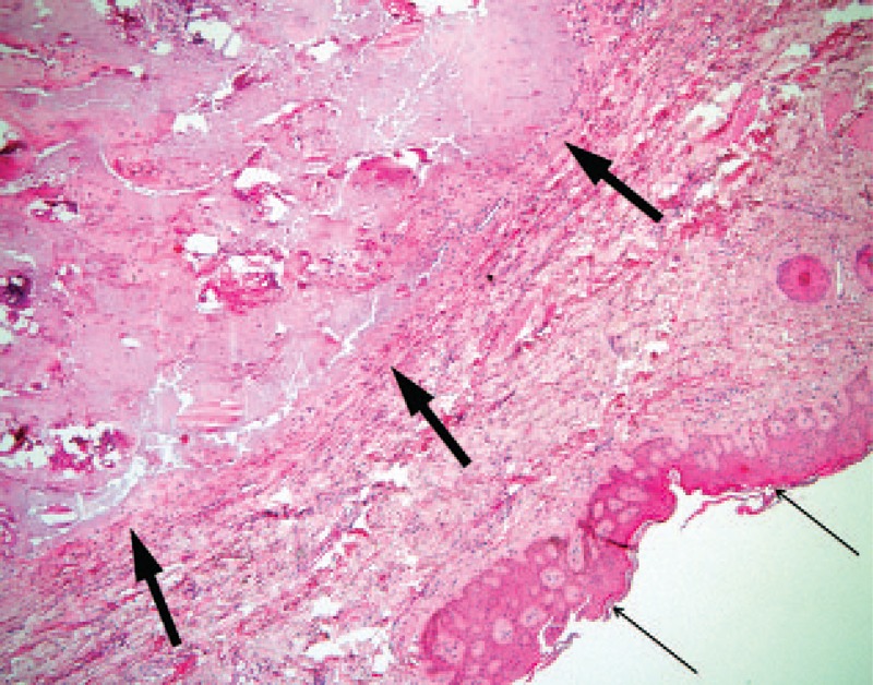
Rationale: Gouty panniculitis, characterised by the deposition of monosodium urate crystals in subcutaneous tissue, is a rare clinical manifestation of gout.
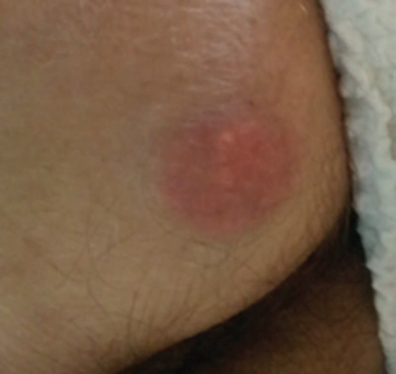
Patient concerns: The case of a 67-year-old man is reported, who presented an erythematous nodule on the upper part of the right buttock suspicious for an abscess. This was in the context of chemotherapy for non-Hodgkin's lymphoma.
Diagnoses: Histopathologic examination demonstrated gouty panniculitis.
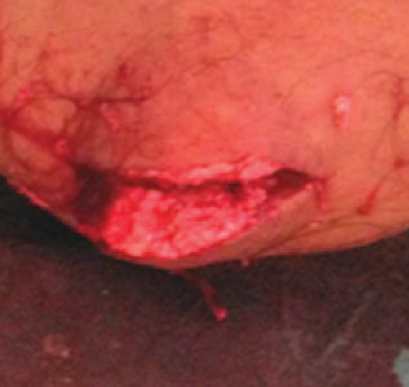
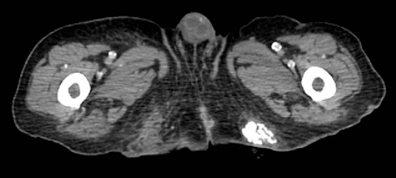
Interventions: Because infection was suspected, an incision was performed. The lesion was found to be densely calcified and friable, without purulent discharge. Therefore, a surgical en-bloc resection was performed.
Outcomes: The wound healed slowly initially due to a combination of malnutrition, chemotherapy and infection. A wound infection with Enterococcus faecium was treated with antibiotic therapy (carbapenem for seven days) and local therapy. At 6-week follow up the wound showed good granulation tissue and was healing well by secondary intention. The patient was instructed to continue anti-hyperuricaemic treatment.
Lessons subsections: In patients known to have long-standing hyperuricaemia and gout with nonspecific subcutaneous erythematous nodules, gouty panniculitis should be considered.
Conflict of interest statement
The authors have no conflicts of interest to disclose.
Figures
Similar articles
-
Gouty panniculitis: a case report and review of the literature.Cutis. 2005 Jul;76(1):54-6. Cutis. 2005. PMID: 16144290 Review.
-
Panniculitis: another clinical expression of gout.Rheumatol Int. 2011 Jun;31(6):831-5. doi: 10.1007/s00296-010-1561-8. Epub 2010 Aug 21. Rheumatol Int. 2011. PMID: 20730459
-
Gouty panniculitis in a 68-year-old man: case report and review of the literature.Int J Dermatol. 2010 Apr;49(4):410-3. doi: 10.1111/j.1365-4632.2010.04283.x. Int J Dermatol. 2010. PMID: 20465696
-
High-frequency ultrasound features in a case of gouty panniculitis.Dermatol Online J. 2017 Jun 15;23(6):13030/qt33j2z14m. Dermatol Online J. 2017. PMID: 28633741
-
Overview of hyperuricaemia and gout.Curr Pharm Des. 2005;11(32):4117-24. doi: 10.2174/138161205774913318. Curr Pharm Des. 2005. PMID: 16375732 Review.
Cited by
-
Auricular Gouty Tophi: A Rare Presentation in an Uncommon Site.Cureus. 2025 Mar 23;17(3):e81035. doi: 10.7759/cureus.81035. eCollection 2025 Mar. Cureus. 2025. PMID: 40264604 Free PMC article.
-
Crystallized but not soluble uric acid elicits pro-inflammatory response in short-term whole blood cultures from healthy men.Sci Rep. 2019 Jul 19;9(1):10513. doi: 10.1038/s41598-019-46935-w. Sci Rep. 2019. PMID: 31324844 Free PMC article.
-
Beyond Medical Treatment: Surgical Treatment of Gout.Curr Rheumatol Rep. 2020 Nov 24;23(1):1. doi: 10.1007/s11926-020-00969-6. Curr Rheumatol Rep. 2020. PMID: 33236200 Review.
-
Multiple Subcutaneous Gouty Tophi Even with Appropriate Medical Treatment: Case Report and Review of Literature.Kans J Med. 2021 Jan 21;14(1):12-16. doi: 10.17161/kjm.vol1414505. eCollection 2021. Kans J Med. 2021. PMID: 33643522 Free PMC article. No abstract available.
References
-
- Marson P, Pasero G. Some historical remarks on microcrystalline arthritis (gout and chondrocalcinosis). Reumatismo 2011;63:199–206. - PubMed
-
- Luk AJ, Simkin PA. Epidemiology of hyperuricemia and gout. Am J Manag Care 2005;11(15 suppl):S435–442. - PubMed
-
- Purohit MB, Purohit TM, Tandon RK. FNAC of gouty tophi—a case report. Indian J Pathol Microbiol 2006;49:42–3. - PubMed
-
- Chhana A, Dalbeth N. The gouty tophus: a review. Curr Rheumatol Rep 2015;17:19. - PubMed
Publication types
MeSH terms
LinkOut - more resources
Full Text Sources
Other Literature Sources
Medical
Research Materials
