. 2017 Apr 19;11(4):e0005548.
doi: 10.1371/journal.pntd.0005548. eCollection 2017 Apr.

The gut microbiota as a modulator of innate immunity during melioidosis

Affiliations

The gut microbiota as a modulator of innate immunity during melioidosis

Jacqueline M Lankelma et al. PLoS Negl Trop Dis. .

Abstract

Background: Melioidosis, caused by the Gram-negative bacterium Burkholderia pseudomallei, is an emerging cause of pneumonia-derived sepsis in the tropics. The gut microbiota supports local mucosal immunity and is increasingly recognized as a protective mediator in host defenses against systemic infection. Here, we aimed to characterize the composition and function of the intestinal microbiota during experimental melioidosis.

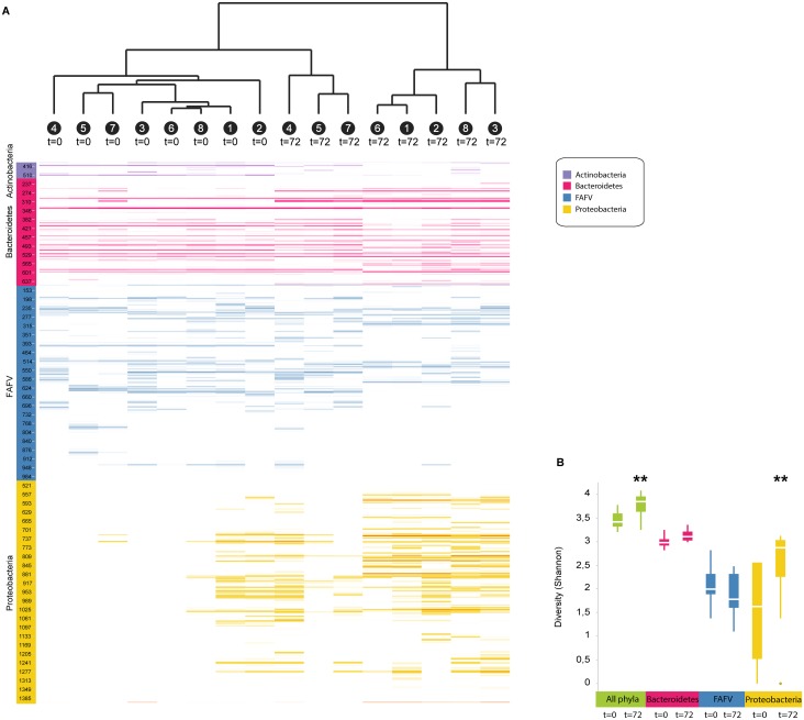
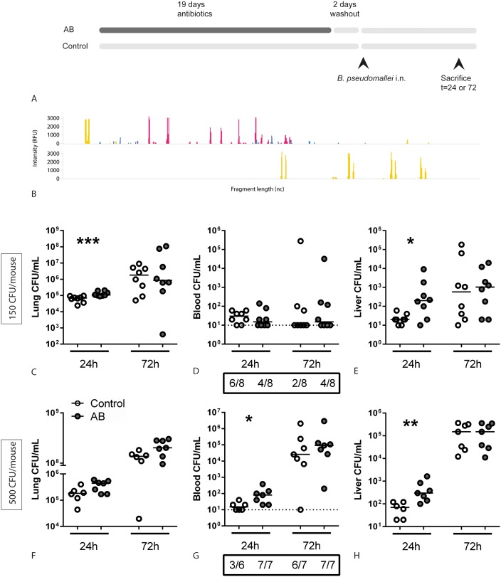
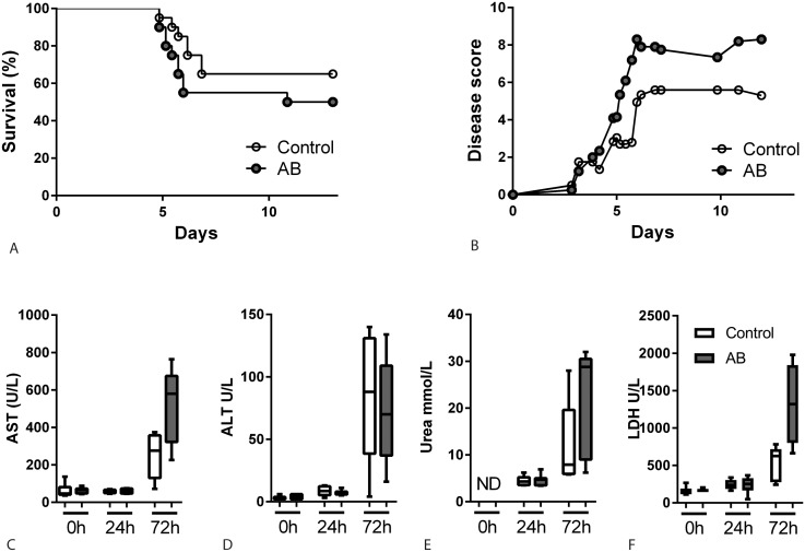
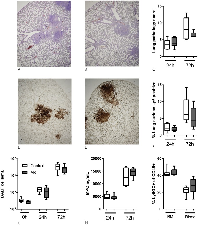
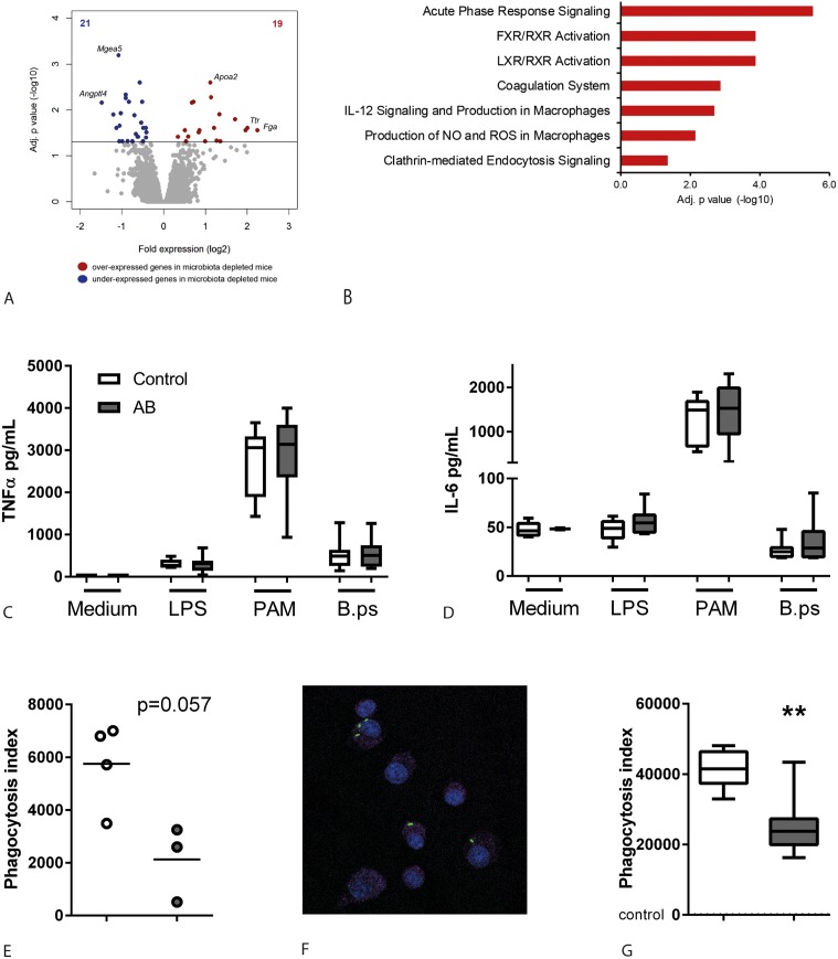
Methodology/principal findings: C57BL/6 mice were infected intranasally with B. pseudomallei and sacrificed at different time points to assess bacterial loads and inflammation. In selected experiments, the gut microbiota was disrupted with broad-spectrum antibiotics prior to inoculation. Fecal bacterial composition was analyzed by means of IS-pro, a 16S-23S interspacer region-based profiling method. A marked shift in fecal bacterial composition was seen in all mice during systemic B. pseudomallei infection with a strong increase in Proteobacteria and decrease in Actinobacteria, with an increase in bacterial diversity. We found enhanced early dissemination of B. pseudomallei and systemic inflammation during experimental melioidosis in microbiota-disrupted mice compared with controls. Whole-genome transcriptional profiling of the lung identified several genes that were differentially expressed between mice with a normal or disrupted intestinal microbiota. Genes involved in acute phase signaling, including macrophage-related signaling pathways were significantly elevated in microbiota disrupted mice. Compared with controls, alveolar macrophages derived from antibiotic pretreated mice showed a diminished capacity to phagocytose B. pseudomallei. This might in part explain the observed protective effect of the gut microbiota in the host defense against pneumonia-derived melioidosis.

Conclusions/significance: Taken together, these data identify the gut microbiota as a potential modulator of innate immunity during B. pseudomallei infection.


Conflict of interest statement

The authors have declared that no competing interests exist.

Figures

Fig 1
Fig 1. Profound changes in fecal microbiota composition during melioidosis.
Fecal pellets were sampled from eight mice before (t = 0) they were infected intranasally with 150 CFU of B. pseudomallei and 72 hours after (t = 72). Microbial composition was analysed by IS-pro, using the number of nucleotides between the genes for ribosomal subunit 16 and 23 in the DNA (interspacer region) of the bacterium as a unique classification characteristic. (A) Clustering analysis, by unweighted pair group method with arithmetic mean (UPGMA) on cosine distances, shows the similarity of samples; individual mice are indicated by a number. Colors represent the most important bacterial phyla (purple, Actinobacteria; red, Bacteroidetes; blue, Firmicutes, Actinobacteria, Fusobacteria, and Verrucomicrobia (FAFV); yellow, Proteobacteria). Length of the interspacer regions in basepairs is indicated on the y-axis; lines indicate the presence of PCR products. Color intensity increases with the presence of PCR product. (B) Diversity of microbial communities before and 72 hours after induction of melioidosis, expressed as Shannon index (green: total bacteria; red: Bacteroidetes; Blue: Firmicutes, Actinobacteria, Fusobacteria, and Verrucomicrobia (FAFV); yellow: Proteobacteria). Data are presented as box- and whisker plots showing the smallest observation, lower quartile, median, upper quartile and largest observation. ** p<0.01 pre- versus post-infection.
Fig 2
Fig 2. Antibiotic pre-treated mice show increased growth and dissemination of B. pseudomallei during experimental melioidosis.
(A) Study design. (B) Before infection, the fecal microbiota of control- and antibiotic treated mice was analysed by IS-pro. Colors represent the most important bacterial phyla (purple, Actinobacteria; red, Bacteroidetes; blue, Firmicutes, Actinobacteria, Fusobacteria, and Verrucomicrobia (FAFV); yellow, Proteobacteria). Length of the interspacer regions in basepairs is indicated on the x-axis; height of the peaks indicates the presence of PCR products. Samples are pooled from eight mice per group; representative of two experiments. Control and antibiotic pre-treated mice were inoculated intranasally with 150 CFU (C-E) or 500 CFU (F-H) B. pseudomallei and sacrificed at the indicated time points. Bacterial loads in lung homogenate (C, F), blood (D, G) and liver homogenate (E, H) are depicted as scatter dot plots with a line at the median. Numbers in the boxes below (D) and (G) indicate the number of positive blood cultures for the total number of mice. White dots represent control mice, grey dots antibiotic treated mice. N = 6–8 mice per group. * p<0.05, ** p<0.01, *** p<0.001 control versus antibiotic treated.
Fig 3
Fig 3. Limited effect of antibiotic induced gut microbiota disruption on survival and organ damage.
Survival (A) and clinical observation score (B) of control (white dots) and antibiotic treated mice (grey dots) after intranasal inoculation with 150 CFU B. pseudomallei (n = 20 mice per group, depicted is the mean). No statistically significant differences were detected. Aspartate aminotranspherase (AST, C), alanine aminotranspherase (ALT, D), urea (E) and lactate dehydrogenase (LDH, F) were measured in plasma after inoculation with 500 CFU B. pseudomallei as markers for liver-, renal- and general damage. Data are presented as box- and whisker plots showing the smallest observation, lower quartile, median, upper quartile and largest observation. White bars represent control mice, grey bars antibiotic treated mice. N = 5–6 samples per group. ND = not detectable.
Fig 4
Fig 4. Antibiotic microbiota disruption does not affect neutrophil influx.
Lungs were obtained at the indicated time points after intranasal inoculation with 500 CFU B. pseudomallei. Paraffin-embedded lung tissue sections were stained with haematoxylin/eosin to score different parameters for pathology (A-B, 2x magnification, representative images). The combined score, given by a blinded pathologist, was not different between the two groups (C). Sections from the same samples were stained for Ly-6GC as a neutrophil marker (representative microphotographs, 2x magnification) (D-E). The percentage Ly-6GC-positive surface of the total lung surface was calculated using ImageJ (F). The number of cells per mL BALF was counted using a Coulter counter (G). Myeloperoxidase was quantified in lung homogenates as a measure for neutrophil degranulation (H). Bone marrow and blood was obtained from naïve control and antibiotic pre-treated mice and using FACS analysis the percentage of Ly6GC+, CD11b+ cells within the viable CD45+ population was determined (I). Data are presented as box- and whisker plots showing the smallest observation, lower quartile, median, upper quartile and largest observation. White bars represent control mice, grey bars antibiotic treated mice. No statistically significant differences were found.
Fig 5
Fig 5. Impaired phagocytosis of B. pseudomallei by alveolar macrophages derived from gut microbiota disrupted mice.
Naïve antibiotic treated- and control mice were sacrificed after the two-day antibiotic washout period and lungs or alveolar macrophages were harvested. (A) Volcano plot depicting significant (multiple comparison adjusted p<0.05) differentially expressed genes in lungs from naïve control and antibiotic pre-treated mice (microbiota disrupted). Red indicates increased expression; blue indicates decreased expression. (B) Significantly enriched canonical signalling pathways in antibiotic treated mice, represented as a bar plot (Ingenuity pathway analysis). (C-D) Alveolar macrophages from naïve control- and antibiotic treated mice were stimulated for 20 hours with medium, LPS 10 ng/mL, PAM3CSK4 1 ug/mL or heat-killed B. pseudomallei 4x10⁶ CFU/mL and TNFα and IL-6 were measured in supernatant. (E) Alveolar macrophages from naïve control- and antibiotic treated mice (each well pooled from two mice) were incubated with 2,5x10⁷ CFU/mL FITC-labeled heat killed B. pseudomallei and their phagocytic index determined via flowcytometry as described in the Methods. (F) Control and antibiotic microbiota disrupted mice were given 5x10⁶ heat killed FITC-labeled B. pseudomallei intranasally and sacrificed after three hours. Cytospins of BALF were stained with PerCP Cy5.5-CD45 and DAPI to assess whether bacteria were located intracellularly (F, representative image). Alveolar macrophages in BALF were analysed by flowcytometry and phagocytic indexes were calculated (G). Data are presented as box- and whisker plots showing the smallest observation, lower quartile, median, upper quartile and largest observation. White bars represent control mice, grey bars antibiotic treated mice. N = 8 mice per group. ** p<0.01 control versus antibiotic treated.

Similar articles

Cited by

References

    1. Wiersinga WJ, Currie BJ, Peacock SJ. Melioidosis. The New England journal of medicine. 2012;367(11):1035–44. Epub 2012/09/14. 10.1056/NEJMra1204699 - DOI - PubMed
    1. Currie BJ. Melioidosis: evolving concepts in epidemiology, pathogenesis, and treatment. Semin Respir Crit Care Med. 2015;36(1):111–25. 10.1055/s-0034-1398389 - DOI - PubMed
    1. Meumann EM, Cheng AC, Ward L, Currie BJ. Clinical features and epidemiology of melioidosis pneumonia: results from a 21-year study and review of the literature. Clinical infectious diseases: an official publication of the Infectious Diseases Society of America. 2012;54(3):362–9. - PMC - PubMed
    1. Birnie E, Wiersinga WJ, Limmathurotsakul D, Grobusch MP. Melioidosis in Africa: should we be looking more closely? Future Microbiol. 2015;10(2):273–81. 10.2217/fmb.14.113 - DOI - PubMed
    1. Limmathurotsakul D, Golding N, Dance DAB, Messina JP, Pigott DM, Moyes CL, et al. Predicted global distribution of Burkholderia pseudomallei and burden of melioidosis. Nature Microbiology. 2016;1:15008. - PubMed

Publication types

MeSH terms

LinkOut - more resources