. 2017 Apr 19;12(4):e0174690.
doi: 10.1371/journal.pone.0174690. eCollection 2017.

Auxotonic to isometric contraction transitioning in a beating heart causes myosin step-size to down shift

Affiliations

Auxotonic to isometric contraction transitioning in a beating heart causes myosin step-size to down shift

Thomas P Burghardt et al. PLoS One. .

Abstract

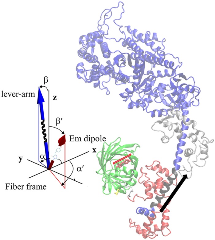
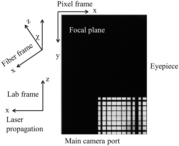
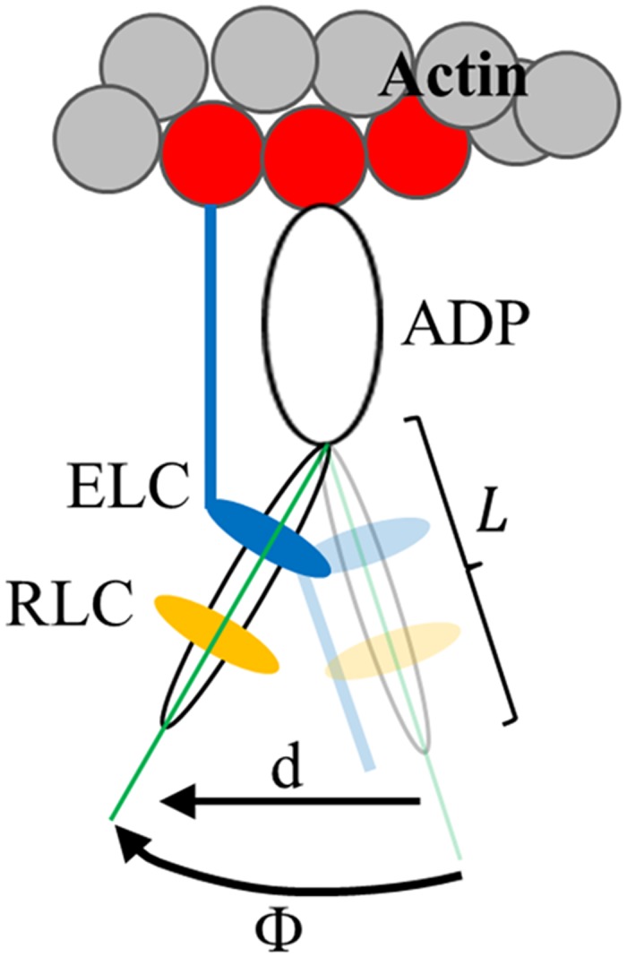
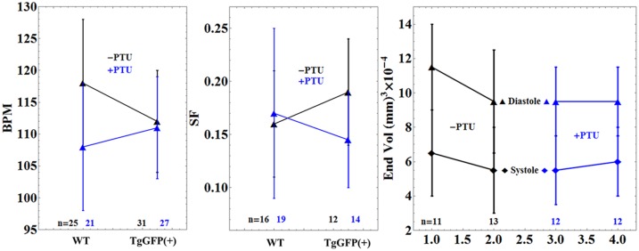
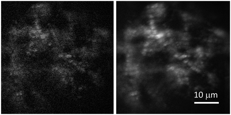
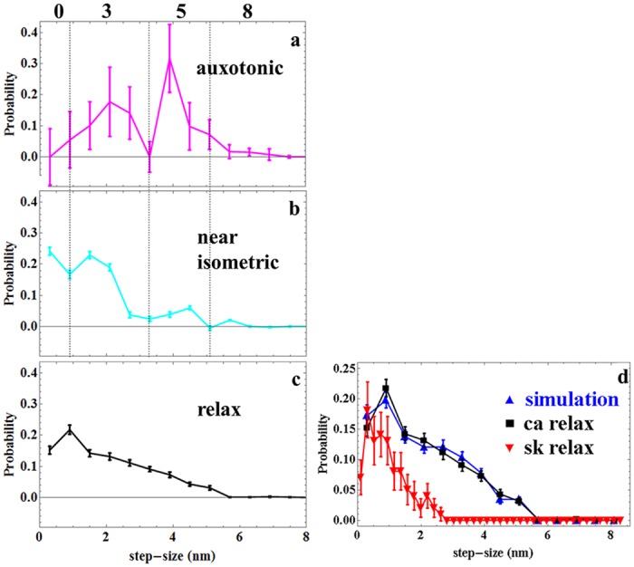
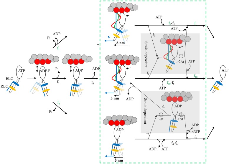
Myosin motors in cardiac ventriculum convert ATP free energy to the work of moving blood volume under pressure. The actin bound motor cyclically rotates its lever-arm/light-chain complex linking motor generated torque to the myosin filament backbone and translating actin against resisting force. Previous research showed that the unloaded in vitro motor is described with high precision by single molecule mechanical characteristics including unitary step-sizes of approximately 3, 5, and 8 nm and their relative step-frequencies of approximately 13, 50, and 37%. The 3 and 8 nm unitary step-sizes are dependent on myosin essential light chain (ELC) N-terminus actin binding. Step-size and step-frequency quantitation specifies in vitro motor function including duty-ratio, power, and strain sensitivity metrics. In vivo, motors integrated into the muscle sarcomere form the more complex and hierarchically functioning muscle machine. The goal of the research reported here is to measure single myosin step-size and step-frequency in vivo to assess how tissue integration impacts motor function. A photoactivatable GFP tags the ventriculum myosin lever-arm/light-chain complex in the beating heart of a live zebrafish embryo. Detected single GFP emission reports time-resolved myosin lever-arm orientation interpreted as step-size and step-frequency providing single myosin mechanical characteristics over the active cycle. Following step-frequency of cardiac ventriculum myosin transitioning from low to high force in relaxed to auxotonic to isometric contraction phases indicates that the imposition of resisting force during contraction causes the motor to down-shift to the 3 nm step-size accounting for >80% of all the steps in the near-isometric phase. At peak force, the ATP initiated actomyosin dissociation is the predominant strain inhibited transition in the native myosin contraction cycle. The proposed model for motor down-shifting and strain sensing involves ELC N-terminus actin binding. Overall, the approach is a unique bottom-up single molecule mechanical characterization of a hierarchically functional native muscle myosin.


Conflict of interest statement

Competing Interests: The authors have declared no competing interests.

Figures

Fig 1
Fig 1. The coordination of the GFP moiety (green) and its emission dipole moment (red arrow) with myosin S1 consisting of a heavy chain (blue and black), ELC (silver), and RLC (red).
The portion of the lever-arm in the heavy chain appearing in black is the α-helix segment associated with the lever-arm orientation and depicted by the black arrow. The insert shows the time-resolved coordinates for GFP chromophore emission dipole moment in red and lever-arm helix in blue corresponding to spherical polar angles (β’,α’) and (β,α) defined relative to a fiber fixed frame.
Fig 2
Fig 2. The fiber frame xz plane in Fig 1 lies in the microscope focal plane and in the lab frame as shown.
Lab and fiber frames are related to each other by a rotation through χ. Images of the heart shown subsequently relate to the lab frame like the image of the test pattern.
Fig 3
Fig 3. Myosin S1 consisting of a heavy chain, ELC (blue), and RLC (yellow) performing a powerstroke with a lever-arm rotation while strongly actin bound.
The arc subtended by, Φ, the angle a single lever-arm helix rotates in sequential images defines a sequence of chords on a circle of diameter L equal to the lever-arm length indicated. Chord length is step-size, d, given by Eq 3. The ELC N-terminal extension, shown actin bound at the end of the powerstroke, plays a major role in the proposed mechanism for strain sensing in the myosin.
Fig 4
Fig 4. The Sypro Ruby stained and immunoblotted SDS-PAGE gels.
The Sypro Ruby gel band intensities establish ZRLC content in a zebrafish adult heart extract relative to a known amount of in vitro expressed HCRLC (panel a). These samples produce calibrated blot intensities for HCRLC and ZRLC under a standardized protein immunoblotting protocol using the RLC antibody as described in Methods and previously [11]. The standards are compared to immunoblots from HCRLC-GFP and ZRLC in WT and TgGFP(+) embryos (panel b) to measure relative fractions of HCRLC-GFP and ZRLC content (ZRLCrep, Eq 1). The relative amount of ZRLC in WT and TgGFP(+) embryo hearts confirmed ZRLC content removed (ZRLCrem, Eq 2) by their comparison to control β-actin expression (panel c).
Fig 5
Fig 5. Comparison of heart rate in Beats Per Minute (BPM), Shortening Fraction (SF), and cyclical ventricle volume changes for WT and TgGFP(+) embryos in the presence (blue) and absence (black) of PTU treatment.
Error bars show standard deviation for n embryos.
Fig 6
Fig 6. A single frame (left) and an averaged image of the heart (right) running at 50–60 bpm.
Fig 7
Fig 7
Panels a-c. In vivo cardiac myosin active cycle from auxotonic (force developing), through near-isometric (maximum force), to detachment (relaxation) phases. Error bars indicate standard deviation of the mean for 15 or 24 cardiac muscle fibers from 8 or 6 embryos and corresponding to 1251 or 1436 single photoactivated myosin coordinates from relaxed and active hearts, respectively. Dotted vertical lines in panels a & b define the boundaries between step-size frequencies computed from areas under the curves for nominal steps of 0, 3, 5, and 8 nm (nominal step-size corresponds to the in vivo measured step-sizes of 0, 2, 4, and 6 nm as indicated on the x-axis legend). Areas at step-size boundaries are split equally between adjoining step-sizes. Panel d. Simulated (blue) and observed (black and same as in panel c) relaxed cardiac (ca) myosin step-size distribution. Simulation is based on dispersion of thick filament structure surmised from the atomic model of Alamo et al. [27] as described in METHODS. In vivo relaxed skeletal (sk) myosin step-size from zebrafish embryo trunk muscle (red) is shown for comparison [31].
Fig 8
Fig 8. Myosin flux through the 4-pathway network contrasting 3 phases of muscle contraction in the beating heart.
Myosin begins and ends detached from actin and with ATP bound in the contraction cycle. The green box with dashed line boundaries group the strong actomyosin bound states. Blue vector v at the end of the myosin lever-arm is positive net force on, and positive velocity of, the thick filament in units where amplitudes are equal. Fluxes through the network, fi, differ depending on contraction phases (values in Table 1). Measured values for fi are in green while computed values are indicated in black. Four pathways cross from beginning to end of the contraction cycle. The top pathway populated by flux f1 executes an 8 nm step-size. The middle pathway populated by flux f3 executes a 3 nm step-size. It releases Pi while weakly actin bound without doing work. The bottom pathway populated by flux f4 is branched and executes 5 or 5+3 nm step-sizes. The branch from the bottom pathway is populated by flux f7 and executes the 5+3 nm step-size. Strain sensitivity is modeled with mechanisms in two subpathways within the shaded regions. The upper mechanism is populated by fluxes f2 and f8 from the 8 and 3 nm steps when the ELC N-terminus binds actin for actomyosin in rigor. The taut (blue line), intermediate (green curve), and slack (red wave) ELC N-terminus for muscle in near-isometric, auxotonic, or unloaded phases have high, modest, or zero strain when net force v is zero in isometric, intermediate in auxotonic, or high in unloaded phase. The linear (blue) actin bound ELC N-terminus is proposed to inhibit ATP binding by lowering active site accessibility for ATP at the small arrow near the myosin head. Inhibited ATP binding extends actomyosin attachment time indicated by the clock icon and quantitated in our single myosin measurements as a 0 length step. The lower mechanism is populated by flux f6 from the 5 nm step with ADP bound. Near-isometric, auxotonic, or unloaded phases have high, intermediate, or zero strain (of an unspecified myosin element) when net force is zero, intermediate, or high. Strain lowers ADP release rate. Short duration ADP rate inhibition flux, f7, leaves to continue with the 3 nm step. Long duration ADP rate inhibition flux, f6 − f7, continues with the 0 length step. For either the ATP accessibility or ADP release rate mediated mechanisms (top or bottom strain sensing mechanisms), low net force inhibits myosin cycling by extending the time myosin is strongly actin bound by >2Δt.

References

    1. Huxley HE. The mechanism of muscular contraction. Science. 1969;164:1356–66. - PubMed
    1. Pant K, Watt J, Greenberg M, Jones M, Szczesna-Cordary D, Moore JR. Removal of the cardiac myosin regulatory light chain increases isometric force production. The FASEB Journal. 2009;23:3571–80. 10.1096/fj.08-126672 - DOI - PMC - PubMed
    1. Burghardt TP, Sikkink LA. Regulatory light chain mutants linked to heart disease modify the cardiac myosin lever-arm. Biochemistry. 2013;52:1249–59. 10.1021/bi301500d - DOI - PMC - PubMed
    1. Lossie J, Köhncke C, Mahmoodzadeh S, Steffen W, Canepari M, Maffei M, et al. Molecular mechanism regulating myosin and cardiac functions by ELC. Biochem Biophys Res Commun. 2014;450(1):464–9. 10.1016/j.bbrc.2014.05.142 - DOI - PubMed
    1. Wang Y, Ajtai K, Kazmierczak K, Szczesna-Cordary D, Burghardt TP. N-terminus of Cardiac Myosin Essential Light Chain Modulates Myosin Step-Size. Biochemistry. 2015;55:186–98. 10.1021/acs.biochem.5b00817 - DOI - PMC - PubMed

MeSH terms

LinkOut - more resources