. 2017 Apr 19;12(4):e0175946.
doi: 10.1371/journal.pone.0175946. eCollection 2017.

Interrelationships of VEL1 and ENV1 in light response and development in Trichoderma reesei

Affiliations

Interrelationships of VEL1 and ENV1 in light response and development in Trichoderma reesei

Hoda Bazafkan et al. PLoS One. .

Abstract

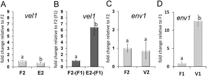
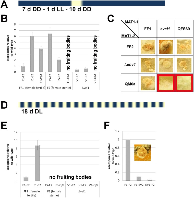
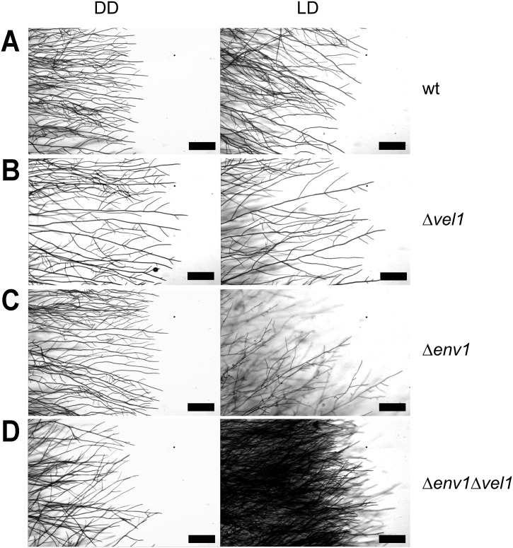
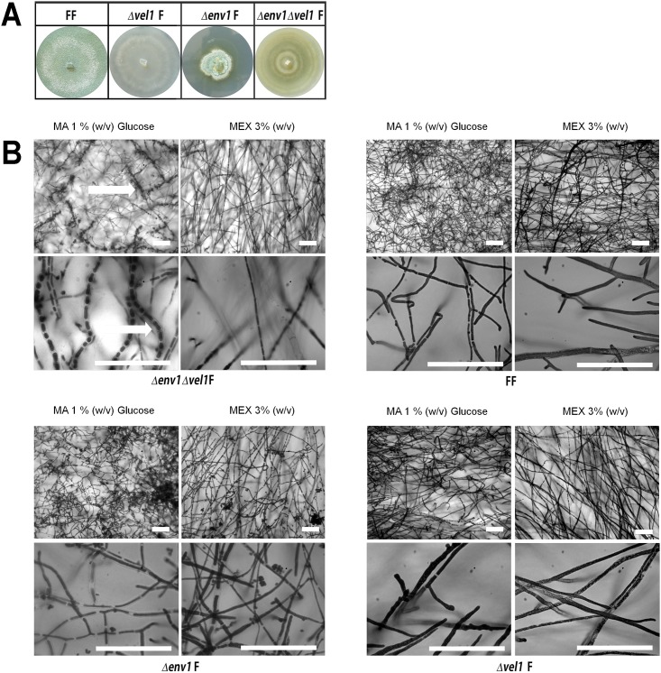
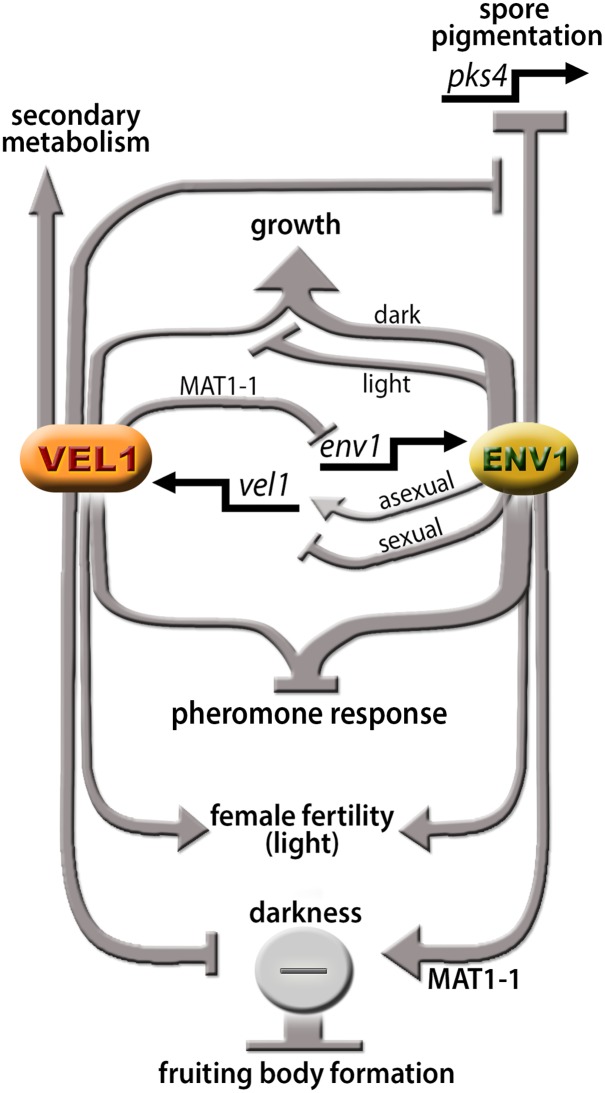
Sexual development is regulated by a complex regulatory mechanism in fungi. For Trichoderma reesei, the light response pathway was shown to impact sexual development, particularly through the photoreceptor ENVOY. Moreover, T. reesei communicates chemically with a potential mating partner in its vicinity, a response which is mediated by the velvet family protein VEL1 and its impact on secondary metabolism. We therefore studied the regulatory interactions of ENV1 and VEL1 with a focus on sexual development. Although individual mutants in both genes are female sterile under standard crossing conditions (light-dark cycles), an altered light regime enabled sexual development, which we found to be due to conditional female sterility of Δenv1, but not Δvel1. Phenotypes of growth and asexual sporulation as well as regulation of the peptide pheromone precursors of double mutants suggested that ENV1 and VEL1 balance positive and negative regulators of these functions. Additionally, VEL1 contributed to the strong deregulation of the pheromone system observed in env1 mutants. Female sterility of Δvel1 was rescued by deletion of env1 in darkness in MAT1-1, indicating a block of sexual development by ENV1 in darkness that is balanced by VEL1 in the wild-type. We conclude that ENV1 and VEL1 exert complementing functions in development of T. reesei. Our results further showed that the different developmental phenotypes of vel1/veA mutants in T. reesei and Aspergillus nidulans are not due to the presence or function of ENV1 in the VELVET regulatory pathway in T. reesei.


Conflict of interest statement

Competing Interests: The authors have declared that no competing interests exist.

Figures

Fig 1
Fig 1. Analysis of mutual regulation of ENV1 and VEL1.
Transcript abundance is shown upon growth on malt extract agar plates at subjective noon as analyzed by qRT-PCR in the female fertile wildtype strain FF1 (“F1”; MAT1-1), (FF2 (“F2”; MAT1-2), in Δenv1F2 (“E2”; MAT1-2) or in Δvel1F2 (“V2”; MAT1-2). (A) Regulation of vel1 transcript abundance by wildtype and in a strain lacking env1 under asexual conditions. (B) Regulation of vel1 transcript abundance under sexual conditions in wildtype F2-(F1) and Δenv1F2 E2-(F1) upon encounter of the female fertile wildtype strain FF1 (“F1”; MAT1-1). (C) Regulation of env1 transcript abundance by VEL1 under asexual conditions in MAT1-2 and (D) in MAT1-1. Pooled samples from several plates and at least two independent biological replicates were considered. Statistical significance of differences was evaluated with the software qbase+ and applying ANOVA analysis with a p-value threshold of p<0.05. Errorbars show standard deviations. Different letters indicate significant differences. For investigation of sexual development, contamination of total RNA of one sample with that of the respective mating partner on the same plate was determined to be below 1.02%.
Fig 2
Fig 2. Fruiting body formation and ascospore discharge under altered light conditions.
(A) Schematic representation of the altered light regime to enable mating of Δenv1F and Δvel1F. (B) Ascospore discharge after 18 days of cultivation under the altered light regime, normalized to the wildtype cross (F1-F2). (C) Fruiting body formation under the altered light conditions. Representative fruiting bodies from 10 replicates cultivated under similar conditions are shown. Red background indicates no fruiting body formation. (D) Schematic representation of light conditions for control cultures under conventional conditions for crossing (daylight, 12:12 cycles). (E) Ascospore discharge after 18 days of cultivation under conventional light-dark cycles, normalized to the wildtype cross (F1-F2). (F) Ascospore discharge of Δenv1Δvel1F double mutants in both mating types after 18 days (light-dark cycles), relative to the wild-type cross (F1-F2). The insert shows a typical fruiting body of a cross of double mutants with wild-type. As Δenv1Δvel1F double mutants are female sterile, they were not able to undergo sexual development with each other. Strains were grown on malt extract medium (3% w/v) at 22°C to enable mating for 18 days under the light conditions described above. Five biological replicates were considered for the analysis and errorbars show standard deviations. Abbreviations: F1: strain FF1, MAT1-1, and F2: strain FF2, MAT1-2 are female fertile derivatives of QM6a obtained by backcrossing [34]; QM: QM6a (MAT1-2); FS: QFS69 (MAT1-1) female sterile derivative of QM6a, sister strain of FF1 and FF2 [34] E2: Δenv1F MAT1-2; V1: Δvel1F MAT1-1; EV1 and EV2: Δenv1Δvel1F double mutants in both mating types.
Fig 3
Fig 3. Microscopic analysis of the phenotype caused by deletion of env1, vel1 or both.
(A) Wildtype QM6a. (B) Vel1 deletion strain. (C) Env1 deletion strain as control (D) Env1 vel1 double mutant. Strains were grown on malt extract agar (MEX) as carbon source in darkness (DD) or daylight (LD, light-dark cycles). Scale bars are equivalent to 200 μm. Lactophenol-blue staining was used to increase contrast in microscopic pictures.
Fig 4
Fig 4. Phenotype of mutants lacking vel1, env1 or both in a female fertile background.
(A) Δvel1 strains are defective in conidiation and Δenv1 mutants in the female fertile background show a similar phenotype as described earlier in female sterile background [15] albeit with a somewhat alleviated defect in conidiation. Double mutants have a mixed phenotype compared to the single mutants. (B) Microscopic analysis using lactophenol-blue staining of Δenv1Δvel1 double mutants and agar-block microscopy. Scale bars are equivalent to 70 μm. Strains were grown on Mandels Andreotti (MA) minimal medium with 1% (w/v) glucose as carbon source or on malt extract agar (MEX) in daylight (light-dark cycles) at 22°C for 10 days.
Fig 5
Fig 5. Analysis of secondary metabolite patterns and transcript levels of pks4 and lxr1.
(A) Secondary metabolite patterns of wildtype (QM6a) and mutant strains are shown during growth under conditions facilitating sexual development (malt extract agar, 3% w/v). Analysis was performed using high performance thin layer chromatography (HPTLC) using at least three biological replicates from pooled samples (3 cultivation plates each), which showed similar results. The figure shows the result from the derivatized HPTLC plate (transmission, visual light). Transcript abundance is shown upon growth on malt extract agar plates at subjective noon as analyzed by qRT-PCR. (B) Regulation of pks4 or (C) lxr1 under asexual conditions in wildtype (F2), Δenv1F (E2), Δvel1F (V2) and Δenv1Δvel1F (EV2). Pooled samples from several plates and at least two independent biological replicates were considered. Statistical significance of differences was evaluated with the software qbase+ and applying ANOVA analysis with a p-value threshold of p<0.05. Errorbars show standard deviations. Different letters indicate significantly different transcript levels.
Fig 6
Fig 6. Analysis of mating and female fertility of Δenv1Δvel1F in light.
Strains were grown on malt extract agar in daylight (light-dark cycles) at 22°C for 30 days. Red background indicates no fruiting body formation and hence abolished mating.
Fig 7
Fig 7. Analysis of mating and female fertility of Δenv1Δvel1F in darkness.
Strains were grown on malt extract agar in constant darkness at 22°C for 30 days. Red background indicates no fruiting body formation and hence abolished mating.
Fig 8
Fig 8. Analysis of transcript levels of pheromone system genes in double mutants.
Transcript abundance is shown upon growth on malt extract agar plates at subjective noon as analyzed by qRT-PCR. (A) Regulation of hpp1 or (B, C) ppg1 or mat1-2-1 (D) under asexual conditions in wildtype (F1, F2) and Δenv1Δvel1F (EV1, EV2). Results for ppg1 are separated into two panels for better comparison. Transcript levels in F1 are 65±11fold higher than in F2. Pooled samples from several plates and at least two independent biological replicates were considered. Statistical significance of differences was evaluated with the software qbase+ and applying ANOVA analysis with a p-value threshold of p<0.05. Errorbars show standard deviations.
Fig 9
Fig 9. Model for the functions and interrelationships of ENV1 and VEL1.
ENV1 and VEL1 show a mutual regulatory interaction at the level of transcription. Both genes contribute to regulation of pheromone response. ENV1 and VEL1 are essential for female fertility in light. In darkness, regulation of fruiting body formation is balanced by the effect of ENV1 and VEL1 on a negative regulator in MAT1-1. If only VEL1 is lacking, the positive effect of ENV1 on the repressor leads to a block of fruiting body formation. Deletion of ENV1 abolishes this positive effect and alleviates the block, which is also in agreement with the only minor function of BLR1 and BLR2 (responsible for induction of env1) in darkness. The largely positive function of VEL1 on secondary metabolism is independent of ENV1, although the specifically negative effect of ENV1 on transcription of pks4 and hence spore/mycelium pigmentation is decreased by VEL1. The effect of ENV1 and VEL1 on growth is light dependent. Arrows indicate a positive function, plungers show a negative effect.

Similar articles

Cited by

References

    1. Glass NL, Schmoll M, Cate JH, Coradetti S. Plant cell wall deconstruction by ascomycete fungi. Annu Rev Microbiol. 2013;67:477–98. 10.1146/annurev-micro-092611-150044 - DOI - PubMed
    1. Schmoll M, Seiboth B, Druzhinina I, Kubicek C. Genomics analysis of biocontrol species and industrial enzyme producers from the genus Trichoderma In: Nowrousian M, editor. Fungal Genomics. The Mycota. XIII Heidelberg, Germany: Springer; 2014. p. 233–65.
    1. Schmoll M, Dattenbock C, Carreras-Villasenor N, Mendoza-Mendoza A, Tisch D, Aleman MI, et al. The genomes of three uneven siblings: footprints of the lifestyles of three Trichoderma species. Microbiol Mol Biol Rev. 2016;80(1):205–327. 10.1128/MMBR.00040-15 - DOI - PMC - PubMed
    1. Schmoll M, Wang TF. Sexual development in Trichoderma In: Wendland J, editor. The Mycota (Vol I): Growth, Differentiation and Sexuality. The Mycota. Switzerland: Springer International Publishing; 2016. p. 457–74.
    1. Seidl V, Seibel C, Kubicek CP, Schmoll M. Sexual development in the industrial workhorse Trichoderma reesei. Proc Natl Acad Sci U S A. 2009;106(33):13909–14. Epub 2009/08/12. 10.1073/pnas.0904936106 - DOI - PMC - PubMed

MeSH terms

LinkOut - more resources