

Comparison of T2*-weighted and QSM contrasts in Parkinson's disease to visualize the STN with MRI
- PMID: 28423027
- PMCID: PMC5397046
- DOI: 10.1371/journal.pone.0176130
Comparison of T2*-weighted and QSM contrasts in Parkinson's disease to visualize the STN with MRI
Abstract
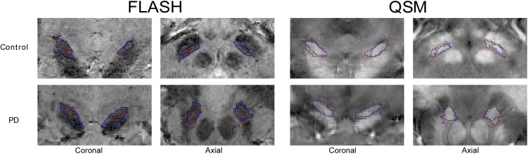
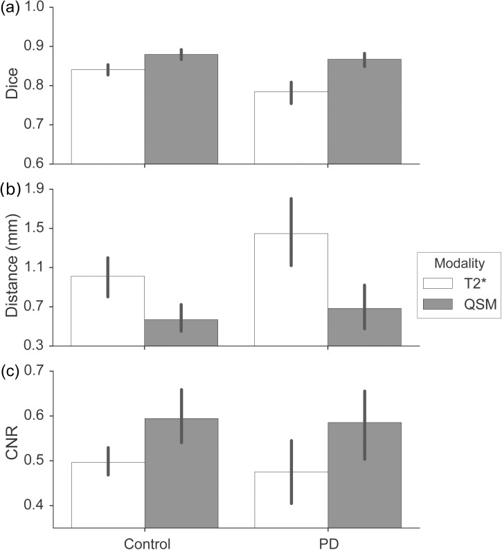
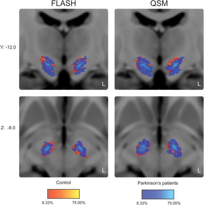
The subthalamic nucleus (STN) plays a crucial role in the surgical treatment of Parkinson's disease (PD). Studies investigating optimal protocols for STN visualization using state of the art magnetic resonance imaging (MRI) techniques have shown that susceptibility weighted images, which display the magnetic susceptibility distribution, yield better results than T1-weighted, T2-weighted, and T2*-weighted contrasts. However, these findings are based on young healthy individuals, and require validation in elderly individuals and persons suffering from PD. Using 7T MRI, the present study set out to investigate which MRI contrasts yielded the best results for STN visualization in 12 PD patients and age-matched healthy controls (HC). We found that STNs were more difficult to delineate in PD as reflected by a lower inter-rater agreement when compared to HCs. No STN size differences were observed between the groups. Analyses of quantitative susceptibility mapping (QSM) images showed a higher inter-rater agreement reflected by increased Dice-coefficients. The location of the center of mass of the STN was not affected by contrast. Overall, contrast-to-noise ratios (CNR) were higher in QSM than in T2*-weighted images. This can at least partially, explain the higher inter-rater agreement in QSM. The current results indicate that the calculation of QSM contrasts contributes to an improved visualization of the entire STN. We conclude that QSM contrast is the preferred choice for the visualization of the STN in persons with PD as well as in aging HC.
Conflict of interest statement
Figures
References
-
- Castrioto A, Lhommee E, Moro E, Krack P. Mood and behavioural effects of subthalamic stimulation in Parkinson’s disease. Lancet Neurol. 2014/02/22. 2014;13: 287–305. doi: 10.1016/S1474-4422(13)70294-1 - DOI - PubMed
-
- Temel Y, Blokland A, Steinbusch HW, Visser-Vandewalle V. The functional role of the subthalamic nucleus in cognitive and limbic circuits. Prog Neurobiol. 2005;76: 393–413. doi: 10.1016/j.pneurobio.2005.09.005 - DOI - PubMed
-
- Richter EO, Hoque T, Halliday W, Lozano AM, Saint-Cyr JA. Determining the position and size of the subthalamic nucleus based on magnetic resonance imaging results in patients with advanced Parkinson disease. J Neurosurg. 2004/03/24. 2004;100: 541–546. - PubMed
-
- Boviatsis EJ, Stavrinou LC, Themistocleous M, Kouyialis AT, Sakas DE. Surgical and hardware complications of deep brain stimulation. A seven-year experience and review of the literature. Acta Neurochir. 2010/07/27. 2010;152: 2053–2062. doi: 10.1007/s00701-010-0749-8 - DOI - PubMed
-
- Chandran AS, Bynevelt M, Lind CRP. Magnetic resonance imaging of the subthalamic nucleus for deep brain stimulation. American Association of Neurological Surgeons; 124: 96–105. doi: 10.3171/2015.1.JNS142066 - DOI - PubMed
MeSH terms
LinkOut - more resources
Full Text Sources
Other Literature Sources
Medical
