. 2017 Apr 19;12(4):e0175223.
doi: 10.1371/journal.pone.0175223. eCollection 2017.

Genetic profiling of putative breast cancer stem cells from malignant pleural effusions

Affiliations

Genetic profiling of putative breast cancer stem cells from malignant pleural effusions

Verena Tiran et al. PLoS One. .

Abstract

A common symptom during late stage breast cancer disease is pleural effusion, which is related to poor prognosis. Malignant cells can be detected in pleural effusions indicating metastatic spread from the primary tumor site. Pleural effusions have been shown to be a useful source for studying metastasis and for isolating cells with putative cancer stem cell (CSC) properties. For the present study, pleural effusion aspirates from 17 metastatic breast cancer patients were processed to propagate CSCs in vitro. Patient-derived aspirates were cultured under sphere forming conditions and isolated primary cultures were further sorted for cancer stem cell subpopulations ALDH1+ and CD44+CD24-/low. Additionally, sphere forming efficiency of CSC and non-CSC subpopulations was determined. In order to genetically characterize the different tumor subpopulations, DNA was isolated from pleural effusions before and after cell sorting, and compared with corresponding DNA copy number profiles from primary tumors or bone metastasis using low-coverage whole genome sequencing (SCNA-seq). In general, unsorted cells had a higher potential to form spheres when compared to CSC subpopulations. In most cases, cell sorting did not yield sufficient cells for copy number analysis. A total of five from nine analyzed unsorted pleura samples (55%) showed aberrant copy number profiles similar to the respective primary tumor. However, most sorted subpopulations showed a balanced profile indicating an insufficient amount of tumor cells and low sensitivity of the sequencing method. Finally, we were able to establish a long term cell culture from one pleural effusion sample, which was characterized in detail. In conclusion, we confirm that pleural effusions are a suitable source for enrichment of putative CSC. However, sequencing based molecular characterization is impeded due to insufficient sensitivity along with a high number of normal contaminating cells, which are masking genetic alterations of rare cancer (stem) cells.


Conflict of interest statement

Competing Interests: This study was funded by the Oesterreichische Nationalbank with the grant number 14001, but the funding source had no influence in study design, data collection and analysis, decision to publish, or preparation of the manuscript. This does not alter our adherence to PLoS One policies on sharing data and materials.

Figures

Fig 1
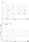
Fig 1. Expression of cancer stem cell markers ALDH1 and CD44+/CD24-/low in malignant pleural effusions of metastatic breast cancer patients.
(A) Aldefluor assay measuring ALDH1 expression in individual pleura samples. (B) FACS dot plots of CD44+/CD24-/low staining. First plots show settings for linage cocktail staining, positive cells were depleted. Both images A and B also include a representative plot of an unstained sample.
Fig 2
Fig 2. Sphere formation assay.
Representative pictures from mammosphere assay. (A) PL11, left sphere from unsorted cells, middle sphere from CD44+/CD24-/low subpopulation, right cells from CD44-/CD24- subpopulation. (B) PL13, left sphere from unsorted cells, middle sphere from ALDH1+ subpopulation, right cells from ALDH1- subpopulation. Scale bar 50 μM. (C) Summary of sphere formation efficiency of all pleura samples with their different subpopulations.
Fig 3
Fig 3. Copy number profiles.
Primary tumors of PL21 (A) and PL24 (E), the cultivated unsorted cells before cell sorting (B) (F), CD44-positive cell fraction of PL21 (C) and the ALDH1+ cell fraction of PL21 (D), respectively, after FACS cell sorting. PL24 long-term cultivation of unsorted spheroids (G). Depicted are segmented log2-ratio plots of the genome. The X-axis indicates the chromosome and Y-axis indicate the log2-ratios. Regions with log2 ratios > 2 indicate gain of chromosomal material and those regions with log2 ratios < 2 indicate loss of chromosomal material.
Fig 4
Fig 4. Characterization of PL24.
(A) Selected copy number plots of PL24. Depicted are segmented log2-ratio plots of chromosomes 1, 6, 17 and 18. (B) Representative picture of spheres in culture. (C) FACS dot plots of PL24 passage 20 left control with DEAB and right Aldefluor assay staining. (D) Immunofluorescent staining of FFPE PL24 spheres I ALDH1 (red) CK (green), II CD44 (red) CK (green), III Ki67 (red) CK (green), IV HEA (red) CK (green), V Vim (red) CK (green), VI Her2neu (green). All slides were counterstained with Dapi (blue). Measuring bar: 100 μm.

References

    1. Siegel RL, Miller K, Jemal A. Cancer statistics. CA Cancer J Clin. 2015;65(1):5–29. 10.3322/caac.21254 - DOI - PubMed
    1. Giuliano A, Hawes D, Ballman K. Association of Occult Metastases in Sentinel Lymph Nodes and Bone Marrow With Survival Among Women With Early-Stage Invasive Breast Cancer. JAMA. 2011;306(4):385–93. 10.1001/jama.2011.1034 - DOI - PMC - PubMed
    1. Balic M, Schwarzenbacher D, Stanzer S, Heitzer E, Auer M, Geigl JB, et al. Genetic and epigenetic analysis of putative breast cancer stem cell models. BMC Cancer. 2013;13. - PMC - PubMed
    1. Dontu G, Al-Hajj M, Abdallah WM, Clarke MF, Wicha MS. Stem cells in normal breast development and breast cancer. Cell Prolif. 2003;36:59–72. - PMC - PubMed
    1. Baccelli I, Trumpp A. The evolving concept of cancer and metastasis stem cells. J Cell Biol. 2012;196(3):281–93. - PMC - PubMed

MeSH terms