. 2017 Apr 18;46(4):621-634.
doi: 10.1016/j.immuni.2017.03.020.

Germ-Cell-Specific Inflammasome Component NLRP14 Negatively Regulates Cytosolic Nucleic Acid Sensing to Promote Fertilization

Affiliations

Germ-Cell-Specific Inflammasome Component NLRP14 Negatively Regulates Cytosolic Nucleic Acid Sensing to Promote Fertilization

Takayuki Abe et al. Immunity. .

Abstract

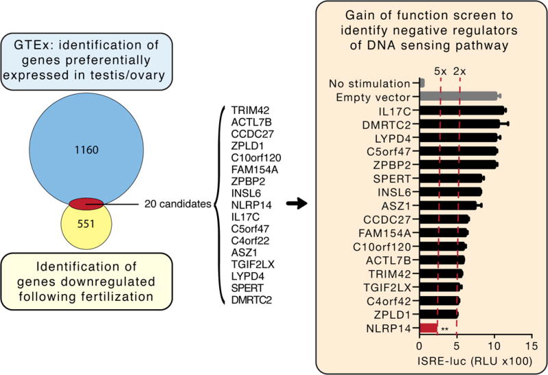
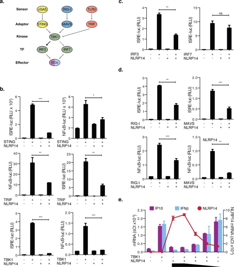
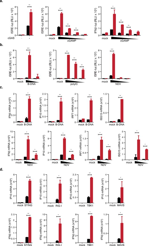
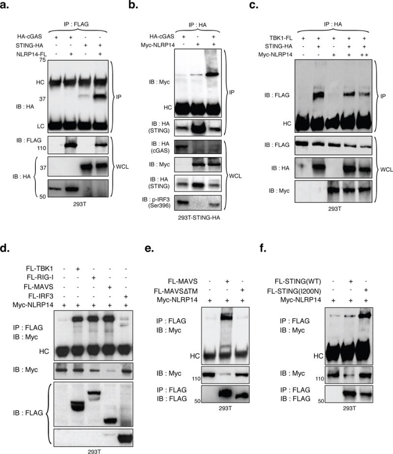
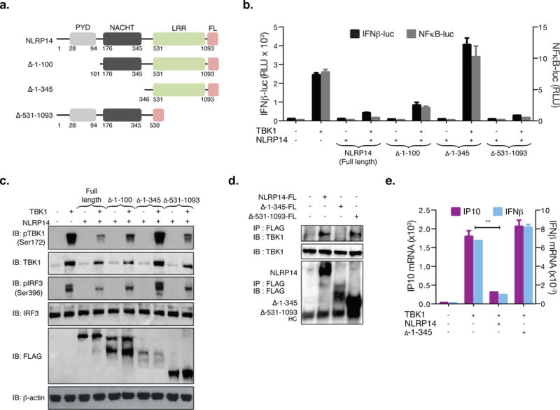
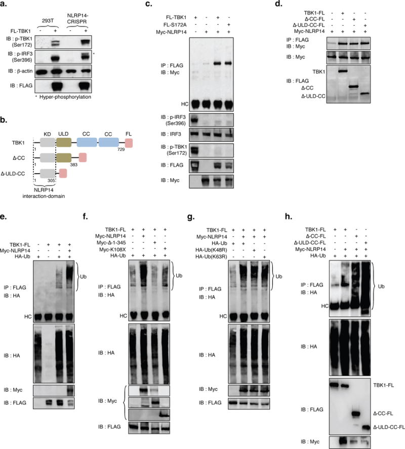
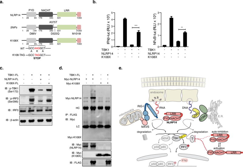
Cytosolic sensing of nucleic acids initiates tightly regulated programs to limit infection. Oocyte fertilization represents a scenario wherein inappropriate responses to exogenous yet non-pathogen-derived nucleic acids would have negative consequences. We hypothesized that germ cells express negative regulators of nucleic acid sensing (NAS) in steady state and applied an integrated data-mining and functional genomics approach to identify a rheostat of DNA and RNA sensing-the inflammasome component NLRP14. We demonstrated that NLRP14 interacted physically with the nucleic acid sensing pathway and targeted TBK1 (TANK binding kinase 1) for ubiquitination and degradation. We further mapped domains in NLRP14 and TBK1 that mediated the inhibitory function. Finally, we identified a human nonsense germline variant associated with male sterility that results in loss of NLRP14 function and hyper-responsiveness to nucleic acids. The discovery points to a mechanism of nucleic acid sensing regulation that may be of particular importance in fertilization.

Keywords: DNA/RNA sensing; RIG-I; STING; TBK1; fertilization; inflammasome; innate immunity; nucleic acid sensing; regulation.


Figures

Figure 1
Figure 1. Data-driven candidate discovery and targeted functional screen identifies putative regulators of cytosolic nucleic acid sensing in germ cells
Left panel: Venn diagram of genes preferentially expressed in testis and/or ovary and down-regulated following oocyte fertilization (see Materials for further description of the discovery method). Of the 20 candidates identified, 17 (listed) were successfully cloned and expressed in 293T cells. Right panel: 293T cells stably expressing STING-HA (293T-STING-HA) were cotransfected with cGAS-Flag and each candidate along with the luciferase gene under control of the ISRE promoter and monitored for luciferase expression 24h post transfection. Shown are relative luciferase units (RLU) normalized to Renilla luciferase expression. Dotted red lines indicate 2- and 5-fold suppression. **P<0.05, Student’s test. Error bars indicate the SD. Data is representative of at least two independent experiments.
Figure 2
Figure 2. NLRP14 suppresses TBK1-dependent innate immunity
(A) Schematic diagram outlining signaling pathway components involved in cytosolic nucleic acid sensing. (B–D) 293T cells were cotransfected with Myc-NLRP14 and RIG-I-Flag, MAVS-Flag, STING-HA, TBK1-Flag, TRIF-Flag, IRF3-Flag, or untagged-IRF7 along with the luciferase gene under control of the ISRE or NFκB promoter. Shown are relative luciferase units (RLU) normalized to Renilla luciferase expression. (E) 293T cells were cotransfected with TBK1-Flag and decreasing amounts of Myc-NLRP14. IP10, IFNβ, and NLRP14 mRNA levels were determined by real-time PCR 24h post transfection. RT-PCR data were normalized to GAPDH mRNA. **P<0.01, Student’s T-test. Error bars indicate SD. Data are representative of at least three independent experiments. (See also Supplemental Figure S1 and S3)
Figure 3
Figure 3. Loss of NLRP14 results in augmentation of immune responses via the cGAS/RIG-I axis
(A) 293T-STING-HA-NLRP14-CRISPR cells were transfected with cGAS-Flag, or decreasing doses of 2′3′-cGAMP (30, 15, 7.5 and 1.5 μg/ml) along with ISRE or IFNβ promoters. Shown are relative luciferase units (RLU) normalized to Renilla luciferase expression. (B) Parental and 293T-NLRP14-CRISPR cells were stimulated with B-DNA (0.06 or 0.03 μg/ml), PolyI:C (25, 12.5, and 2.5 μg/ml), or infected with NDV-GFP (moi of 20, 4, or 0.8). 24h after treatment, levels of ISRE inducing cytokines in supernatants were determined using 293T-ISRE-B8 cells. (C) Rhodamine-labeled B-DNA (indicated as B-DNA Rho) was transfected into parental (WT) and 293T-NLRP14-CRISPR (KO) cells, and observed by fluorescence microscopy. (D) Parental and 293T-NLRP14-CRISPR cells were stimulated with indicated ligands (0.06 μg/ml of B-DNA and NDV-GFP at the m.o.i of 10), and levels of IP10, IFNβ, IRF7 and ISG15 mRNA were determined by RT-PCR 6h post transfection of B-DNA and 15h post NDV inoculation. (E) Parental and 293T-NLRP14-CRISPR cells were transfected with RIG-I-Flag, MAVS-Flag, TBK1-Flag, or STING-HA, and IP10 and IFNβ mRNA levels were determined by RT-PCR at 24h post transfection. RT-PCR data were normalized to GAPDH mRNA. **P<0.01, ***P<0.001, Student’s T-test. Error bars indicate SD. Data are representative of at least three independent experiments. Black and red bars indicate parental and CRISPR-targeted cells, respectively. (See also Supplemental Figures S2 and S4)
Figure 4
Figure 4. NLRP14 interacts with components of cytosolic nucleic acid sensing machinery
(A) NLRP14-Flag was coexpressed with HA-cGAS or STING-HA in 293T cells and subjected to immunoprecipitation (IP) and immunoblotting (IB) using appropriate antibodies. (B) Myc-NLRP14 was coexpressed with or without HA-cGAS in 293T-STING-HA cells and subjected to IP and IB using the appropriate antibodies. (C) The effect of NLRP14 on TBK1-STING interaction was examined in the context of 1μg (+) or 2μg (++) Myc-NLRP14 expression plasmid. Myc-NLRP14 was coexpressed with (D) RIG-I-Flag, MAVS-Flag, TBK1-Flag, or IRF3-Flag (E) FL-MAVS or FL-MAVSΔTM (F) FL-STING or FL-STING(I200N) in 293T cells and monitored for interactions by IP followed by IB using appropriate antibodies. WCL, HC and LC indicate whole cell lysates, Heavy-Chain, and Light-Chain, respectively. (See also Supplemental Figure S6)
Figure 5
Figure 5. N-terminal PYD and NACHT domains mediate NLRP14 function
(A) Schematic of human NLRP14 and deletion mutants generated. Pyrin domain (PYD); NAIP, CIITA, HET-E, and TP1 domain (NACHT); Leucine rich repeat (LRR). 293T cells were cotransfected with TBK1 and NLRP14 variants along with IFNβ-luc or NFκB-luc and monitored for promoter activity (RLU; B) or subjected to immunoblotting with appropriate antibodies (C). (D) 293T cells were transfected with full length, Δ1-345 or Δ531-1093 NLRP14 and interaction with endogenous TBK1 monitored by immunoprecipitation 48h post transfection. (E) 293T cells were cotransfected with TBK1 and NLRP14 variants and IP10 and IFNβ mRNA levels were determined by RT-PCR 24h post transfection. **P<0.01, Student’s T-test. Error bars indicate SD. Data are representative of at least two independent experiments. (See also Supplemental Figure S6)
Figure 6
Figure 6. NLRP14 dampens TBK1-mediated signaling through interactions with TBK1-kinase domain (KD) and induction of TBK1 ubiquitination
(A) Parental and 293T-NLRP14-CRISPR cells were transfected with TBK1-Flag and 24h later monitored for TBK1 expression and phosphorylation (S172), phosphorylation of IRF3 (S396), and β-actin using appropriate antibodies. Asterisks indicates hyper-phosphorylation of IRF3. (B) Schematic of human TBK1 and deletion mutants generated. Kinase domain (KD); ubiquitin-like domain (ULD); coiled-coil domain (CC). (C, D) Immunoprecipitation and immunoblot of 293T cell extracts transfected with various combinations (noted above the lanes) of NLRP14 and TBK1 mutants using the appropriate antibodies. (E–H) Immunoprecipitation and immunoblot of 293T cell extracts transfected with various combinations (noted above lanes) of NLRP14 variants, TBK1 variants, and linked ubiquitin. (I) Schematic of human TBK1 K670R (constitutively active) variant generated. (J) ISRE-luc and NFκB-luc activity in cells transfected with combinations of TBK1-FL, myc-NLRP14, and K670R TBK1 mutant. Shown are relative luciferase units (RLU) normalized to Renilla luciferase expression. **P<0.01, Student’s T-test. Error bars indicate SD. Data are representative of at least two independent experiments. (See also Supplemental Figure S6)
Figure 7
Figure 7. Naturally occurring SNP associated with male sterility results in loss of NLRP14 function
(A) Schematic of human NLRP14 and location of SNPs identified in men with spermatogenic failure. (B) 293T cells were transfected with combinations (noted above lanes) of NLRP14 (WT and K108X mutant), TBK1 and (where noted) IFNβ-luc or NFκB-luc and monitored for luciferase activity (B; shown are RLU normalized to Renilla luciferase expression), phosphorylation of TBK1 and IRF3 (C), and TBK1-NLRP14 interactions by immunoprecipitation and immunoblotting (D). (E) Summary model of NLRP14-mediated suppression of cytosolic nucleic acid sensing. **P<0.01, ***P<0.001, Student’s T-test. Error bars indicate SD. Data are representative of at least two independent experiments. (See also Supplemental Figure S7)

Comment in

References

    1. Barber GN. STING: infection, inflammation and cancer. Nature reviews. Immunology. 2015;15:760–770. - PMC - PubMed
    1. Baril M, Racine ME, Penin F, Lamarre D. MAVS dimer is a crucial signaling component of innate immunity and the target of hepatitis C virus NS3/4A protease. Journal of virology. 2009;83:1299–1311. - PMC - PubMed
    1. Cai X, Chiu YH, Chen ZJ. The cGAS-cGAMP-STING pathway of cytosolic DNA sensing and signaling. Molecular cell. 2014;54:289–296. - PubMed
    1. Chen GY. Role of Nlrp6 and Nlrp12 in the maintenance of intestinal homeostasis. European journal of immunology. 2014;44:321–327. - PMC - PubMed
    1. Consortium GT. The Genotype-Tissue Expression (GTEx) project. Nature genetics. 2013;45:580–585. - PMC - PubMed

MeSH terms