

MDI 301, a synthetic retinoid, depressed levels of matrix metalloproteinases and oxidative stress in diabetic dermal fibroblasts
- PMID: 28423369
- PMCID: PMC5546448
- DOI: 10.18632/oncotarget.16803
MDI 301, a synthetic retinoid, depressed levels of matrix metalloproteinases and oxidative stress in diabetic dermal fibroblasts
Abstract
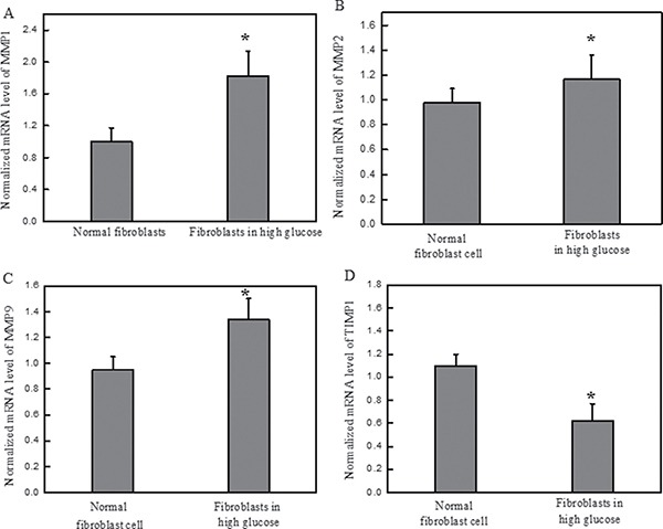
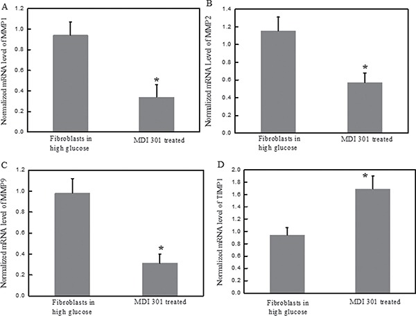
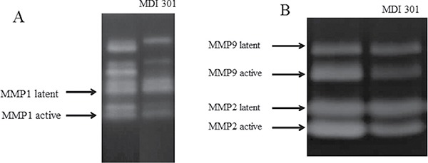
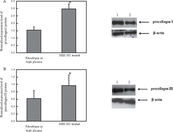
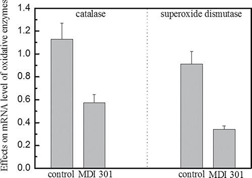
Diabetic foot ulcerations could result in serious consequences such as amputations. The up-regulation of matrix metalloproteinases and down-regulation of TIMP1 were remarked as distinctive biological characteristics in the diabetic dermal fibroblast. The current study was performed in order to clarify the effect of high glucose on formation of diabetic dermal fibroblast cell. In addition, the effect of MDI 301 on ameliorating diabetic fibroblasts was investigated in this study. The mRNA and protein expression levels of MMPs, TIMP1 and catalase were evaluated against fibroblasts treated with high glucose (30 mM) using qRT-PCR, western blotting and zymography assays. Methods were also employed for investigating the biological effects of MDI 301 on high glucose-induced diabetic fibroblasts. In this study, we found that the unbalance of oxidative stress induced by high glucose concentration play an important role in the formation of diabetic dermal fibroblast from normal cells. In addition, MDI 301, a picolinic acid-substituted ester of 9-cis retinoic acid was employed in this study in order to ameliorate symptoms on diabetic dermal fibroblast induced by high glucose concentration. We found MDI 301 alleviate the effects of high glucose-induced skin damage by balancing the oxidative stress and regulating the MMPs and TIMP1 levels. Our finding indicated that MDI 301 offers the potential for repairing the faulty skin function arising from diabetes.
Keywords: MMPs; TIMP1; diabetic fibroblast cell; high glucose; retinoid acid.
Conflict of interest statement
None.
Figures

Similar articles
-
Effects of a synthetic retinoid on skin structure, matrix metalloproteinases, and procollagen in healthy and high-risk subjects with diabetes.J Diabetes Complications. 2011 Nov-Dec;25(6):398-404. doi: 10.1016/j.jdiacomp.2011.10.002. Epub 2011 Nov 4. J Diabetes Complications. 2011. PMID: 22055260 Free PMC article.
-
Myricetin, a potent natural agent for treatment of diabetic skin damage by modulating TIMP/MMPs balance and oxidative stress.Oncotarget. 2016 Nov 1;7(44):71754-71760. doi: 10.18632/oncotarget.12330. Oncotarget. 2016. PMID: 27765936 Free PMC article.
-
23-Hydroxytormentic acid protects human dermal fibroblasts by attenuating UVA-induced oxidative stress.Photodermatol Photoimmunol Photomed. 2017 Mar;33(2):92-100. doi: 10.1111/phpp.12294. Epub 2017 Feb 9. Photodermatol Photoimmunol Photomed. 2017. PMID: 28106292
-
Increased levels of catalase and cathepsin V/L2 but decreased TIMP-1 in keratoconus corneas: evidence that oxidative stress plays a role in this disorder.Invest Ophthalmol Vis Sci. 2005 Mar;46(3):823-32. doi: 10.1167/iovs.04-0549. Invest Ophthalmol Vis Sci. 2005. PMID: 15728537
-
The role of retinoids in the prevention and repair of aged and photoaged skin.Clin Exp Dermatol. 2001 Oct;26(7):613-8. doi: 10.1046/j.1365-2230.2001.00892.x. Clin Exp Dermatol. 2001. PMID: 11696066 Review.
Cited by
-
Oxidative stress in vascular surgical diseases: mechanisms, impacts and therapeutic perspectives.Front Pharmacol. 2025 Apr 9;16:1527684. doi: 10.3389/fphar.2025.1527684. eCollection 2025. Front Pharmacol. 2025. PMID: 40271068 Free PMC article. Review.
-
Evaluation of Gallic Acid-Coated Gold Nanoparticles as an Anti-Aging Ingredient.Pharmaceuticals (Basel). 2021 Oct 22;14(11):1071. doi: 10.3390/ph14111071. Pharmaceuticals (Basel). 2021. PMID: 34832853 Free PMC article.
-
Exploring the influence of growth factors in diabetic foot: A comprehensive bibliometric analysis.Medicine (Baltimore). 2025 Aug 1;104(31):e42716. doi: 10.1097/MD.0000000000042716. Medicine (Baltimore). 2025. PMID: 40760573 Free PMC article.
References
-
- Ulbrecht JS, Cavanagh PR, Caputo GM. Foot problems in diabetes: an overview. Clin Infect Dis. 2004;39:S73–82. - PubMed
-
- Boyko EJ, Ahroni JH, Cohen V, Nelson KM, Heagerty PJ. Prediction of diabetic foot ulcer occurrence using commonly available clinical information: the Seattle Diabetic Foot Study. Diabetes Care. 2006;29:1202–1207. - PubMed
-
- Trengove NJ, Stacey MC, MacAuley S, Bennett N, Gibson J, Burslem F, Murphy G, Schultz G. Analysis of the acute and chronic wound environments: the role of proteases and their inhibitors. Wound Repair Regen. 1999;7:442–452. - PubMed
-
- Lobmann R, Zemlin C, Motzkau M, Reschke K, Lehnert H. Expression of matrix metalloproteinases and growth factors in diabetic foot wounds treated with a protease absorbent dressing. J Diabetes Complications. 2006;20:329–335. - PubMed
MeSH terms
Substances
LinkOut - more resources
Full Text Sources
Other Literature Sources
Medical
Research Materials
Miscellaneous
