. 2017 Mar 28;8(13):21001-21014.
doi: 10.18632/oncotarget.15471.

The PIAS3-Smurf2 sumoylation pathway suppresses breast cancer organoid invasiveness

Affiliations

The PIAS3-Smurf2 sumoylation pathway suppresses breast cancer organoid invasiveness

Amrita Singh Chandhoke et al. Oncotarget. .

Abstract

Tumor metastasis profoundly reduces the survival of breast cancer patients, but the mechanisms underlying breast cancer invasiveness and metastasis are incompletely understood. Here, we report that the E3 ubiquitin ligase Smurf2 acts in a sumoylation-dependent manner to suppress the invasive behavior of MDA-MB-231 human breast cancer cell-derived organoids. We also find that the SUMO E3 ligase PIAS3 inhibits the invasive growth of breast cancer cell-derived organoids. In mechanistic studies, PIAS3 maintains breast cancer organoids in a non-invasive state via sumoylation of Smurf2. Importantly, the E3 ubiquitin ligase activity is required for sumoylated Smurf2 to suppress the invasive growth of breast cancer-cell derived organoids. Collectively, our findings define a novel role for the PIAS3-Smurf2 sumoylation pathway in the suppression of breast cancer cell invasiveness. These findings lay the foundation for the development of novel biomarkers and targeted therapeutic approaches in breast cancer.

Keywords: breast cancer; invasion; sumoylation.


Conflict of interest statement

CONFLICTS OF INTEREST

None.

Figures

Figure 1
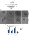
Figure 1. Knockdown of Smurf2 enhances TGFβ-induced disorganization of MDA-MB-231 cancer cell-derived organoids
(A) Lysates of MDA-MB-231 cells transfected with an RNAi plasmid encoding short hairpin RNAs (shRNAs) targeting one of two unique sequences in Smurf2 (Smurf2i-1 or Smurf2i-2), added separately or as a pool (Smurf2i), or the corresponding U6 RNAi plasmid, were immunoblotted with the Smurf2 or actin antibody, the latter serving as loading control. (B) Quantitative analysis of endogenous Smurf2 abundance in lysates of MDA-MB-231 cells transfected as in (A) Bar graph represents the mean ± S.E.M of actin-normalized endogenous Smurf2 abundance expressed relative to the vector control from three independent experiments. Each of the two Smurf2 RNAi plasmids alone or in combination significantly reduced the levels of endogenous Smurf2 in MDA-MB-231 cells. (C) Representative micrographs of 10-day live untreated or TGFβ-incubated three-dimensional organoids derived from MDA-MB-231 cells transfected with an RNAi control vector or a pool of Smurf2i-1 and Smurf2i-2 (Smurf2i) plasmids as described in (A). (D) Quantification of number of invasive MDA-MB-231 cell-derived organoids transfected and treated as in (C). Bar graph represents the mean ± S.E.M of proportion (%) of invasive organoids from three independent experiments including the one in C. TGFβ promotes the invasive behavior of MDA-MB-231 cell-derived organoids. Knockdown of Smurf2 by RNAi also promoted the invasive organoid growth even in the absence of TGFβ. (E) Representative micrographs of 10-day live untreated or TGFβ-incubated organoids derived from MDA-MB-231 cells transfected with control vector alone, or Smurf2i-1 and Smurf2i-2, together with the plasmid expressing Smurf2-r1 and Smurf2-r2, respectively, or with a mammalian expression vector control. (F) Quantification of number of invasive MDA-MB-231 cell-derived organoids transfected and treated as in (E) Bar graph represents the mean ± S.E.M of proportion (%) of invasive organoids from three independent experiments including the one in (E) Smurf2-r1and Smurf2-r2, respectively, reversed Smurf2i-1 and Smurf2i-2-induced invasive behavior of MDA-MB-231 cell-derived organoids in absence and presence of TGFβ. In addition, Smurf2-r1 or Smurf2-r2 coexpression with Smurf2 RNAi suppressed the ability of TGFβ to induce invasive behavior of organoids relative to vector control cells. Significant difference, ANOVA: ***p ≤ 0.001. Scale bar = 50 μm. Mr refers to relative Molecular Mass.
Figure 2
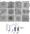
Figure 2. Smurf2 suppresses TGFβ–induced invasive growth of MDA-MB-231 cell-derived organoids in a sumoylation-dependent manner
(A) Lysates of MDA-MB-231 cells expressing wild type Smurf2, Smurf2KdR, or SUMO-Smurf2, or transfected with the control vector, were immunoblotted with the Smurf2 or actin antibody, the latter serving as loading control. (B) Representative micrographs of 10-day live untreated or TGFβ-incubated three-dimensional organoids derived from different MDA-MB-231 cells as in (A). (C) Quantification of number of invasive MDA-MB-231 cell-derived organoids transfected and treated as in A. Bar graph represents the mean ± S.E.M of the proportion (%) of invasive organoids from three independent experiments including the one in (A) Smurf2 and SUMO-Smurf2 suppressed the ability of TGFβ to induce invasive growth in MDA-MB-231 cells. In contrast, Smurf2KdR promoted the invasive growth of breast cancer cell-derived organoids even in the absence of TGFβ. Significant difference, ANOVA: **p ≤ 0.01, ***p ≤ 0.001. Student t-test: #p ≤ 0.05. Scale bar = 50 μm. Mr refers to relative Molecular Mass.
Figure 3
Figure 3. Knockdown of PIAS3 disrupts MDA-MB-231 breast cancer cell-derived organoids
(A) FLAG, PIAS3 or actin immunoblots of lysates of MDA-MB-231 cells cotransfected with a plasmid encoding FLAG-tagged PIAS3 protein, together with an RNAi plasmid encoding short hairpin RNAs (shRNAs) targeting one of two unique sequences in PIAS3, alone or together, or the corresponding U6 RNAi plasmid. Actin served as loading control. The immunoblots are from an experiment that was repeated three independent times with similar results. Expression of PIAS3 shRNA-1 (PIAS3i-1), PIAS3 shRNA-2 (PIAS3i-2) alone or together reduced the abundance of exogenous PIAS3 by more than 90%. (B) PIAS3 and actin immunoblots of lysates of MDA-MB-231 cells transfected with RNAi plasmids encoding short hairpin RNAs (shRNAs) targeting one of two unique sequences in PIAS3 (PIAS3i-1 or PIAS3i-2), added separately or together, or transfected with U6 RNAi plasmid. Actin served as loading control. The immunoblots are from an experiment that was repeated two independent times with similar results. Expression of PIAS3 shRNA-1 (PIAS3i-1), PIAS3 shRNA-2 (PIAS3i-2) alone or together reduced the abundance of endogenous PIAS3 by more than 60%. (C) Representative micrographs of 10-day-old live untreated or TGFβ-incubated three-dimensional organoids derived from MDA-MB-231 cells transfected with an RNAi control vector, PIAS3i-1 plasmid, PIAS3i-2 plasmid or a pool of PIAS3i-1 and PIAS3i-2 (PIAS3i) plasmids as described in (B). (D) Quantification of number of invasive MDA-MB-231 cell-derived organoids transfected and treated as in C. Bar graph represents the mean ± S.E.M of proportion (%) of invasive organoids from four independent experiments including the one in (D). Endogenous PIAS3 knockdown by PIAS3 shRNA-1, PIAS3 shRNA-2, alone or together promoted invasive growth of MDA-MB-231-derived organoids. Significant difference, ANOVA: ***p ≤ 0.001. Scale bar = 50 μm. Mr refers to relative Molecular Mass.
Figure 4
Figure 4. PIAS3 suppresses TGFβ-induced invasive growth of MDA-MB-231 breast cancer cell-derived organoids in a SUMO E3 ligase activity-dependent manner
(A) PIAS3 and actin immunoblots of lysate of MDA-MB-231 cells expressing FLAG-tagged PIAS3 or the SUMO E3 ligase inactive PIAS3CS, or transfected with the control vector. Actin served as a loading control. (B) Representative micrographs of 10-day live untreated or TGFβ-incubated three-dimensional organoids derived from MDA-MB-231 cells transfected as in (A). (C) Quantification of number of invasive MDA-MB-231 cell-derived organoids transfected and treated as in A. Bar graph represents the mean ± S.E.M of proportion (%) of invasive organoids from seven independent experiments including the one in A. PIAS3 significantly suppressed the ability of TGFβ to induce invasive growth in MDA-MB-231 cell-derived organoids. Interestingly, PIAS3CS promoted the invasive growth of MDA-MB-231 cell-derived organoids even in the absence of TGFβ addition. Significant difference, ANOVA: **p ≤ 0.01, *p ≤ 0.05. Scale bar = 50 μm. Mr refers to relative Molecular Mass.
Figure 5
Figure 5. Sumoylation-defective Smurf2 blocks PIAS3 suppression of invasive growth of MDA-MB-231 breast cancer cell-derived organoids
(A) Representative DIC images of 10-day-old live untreated, TGFβ-treated, or KI-treated three-dimensional multicellular structures from MDA-MB-231 cells expressing wild type PIAS3, the SUMO loss of function Smurf2KdR, alone or together, or transected with the vector control. KI refers to the TGFβ type I receptor kinase inhibitor SB431542. (B) Quantification of number of invasive untreated or TGFβ -treated MDA-MB-231 cell-derived organoids transfected as in A. Bar graph represents the mean ± S.E.M. of the proportion (%) of invasive organoids from three independent experiments including the one in A. Smurf2KdR reversed the ability of PIAS3 to suppress TGFβ-induced invasive growth of MDA-MB-231 cell-derived organoids in absence and presence of TGFβ. Smurf2KdR-induced invasive growth of MDA-MB-231 cell-derived organoids is reversed by incubation of the three-dimensional cultures with 10 μM KI. Significant difference, ANOVA: **p ≤ 0.01 and ***p ≤ 0.001. Scale bar = 50 μm.
Figure 6
Figure 6. SUMO-Smurf2 reverses PIAS3CS to induce invasive growth of MDA-MB-231 breast cancer cell-derived organoids
(A) Representative DIC images of 10-day-old live untreated, TGFβ-treated, or KI-treated three-dimensional multicellular structures from MDA-MB-231 cells expressing the SUMO E3 ligase inactive PIAS3CS, the SUMO gain-of-function SUMO-Smurf2, alone or together, or transfected with the vector control. KI refers to the TGFβ type I receptor kinase inhibitor SB431542. (B) Quantification of invasive MDA-MB-231 cell-derived organoids transfected as in A and either left untreated or incubated with TGFβ. Bar graph represents the mean ± S.E.M. of the proportion (%) of invasive organoids from three independent experiments including the one in (A). SUMO-Smurf2 reversed the ability of PIAS3CS to promote TGFβ-induced invasive growth of MDA-MB-231 cell-derived spheroids both in absence and presence of TGFβ. PIAS3CS-induced invasive growth of MDA-MB-231 cell-derived organoids is reversed by incubation of the three-dimensional cultures with 10 μM of KI. Significant difference, ANOVA: **p ≤ 0.01 and ***p ≤ 0.001. Scale bar = 50 μm
Figure 7
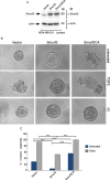
Figure 7. E3 ubiquitin ligase activity is required for Smurf2 to suppress TGFβ-induced invasive growth of MDA-MB-231 cell-derived organoids
(A) Lysates of MDA-MB-231 cells expressing wild type Smurf2 or the ubiquitin E3 ligase inactive Smurf2CA in which Cysteine 716 is mutated to alanine, or transfected with the control vector, were immunoblotted with the MYC or actin antibody, the latter serving as loading control. (B) Representative micrographs of 10-day live untreated, TGFβ incubated or KI-incubated three-dimensional organoids derived from different MDA-MB-231 cells as in (A). (C) Quantification of number of invasive MDA-MB-231 cell-derived organoids transfected and left untreated or incubated with TGFβ are shown. Bar graph represents the mean ± S.E.M of the proportion (%) of invasive organoids from seven independent experiments including the one in (A). Smurf2 suppressed the ability of TGFβ to induce invasive growth in MDA-MB-231 cell-derived organoids. In contrast, Smurf2CA promoted the invasive growth of breast cancer cell-derived organoids even in the absence of TGFβ. Significant difference, ANOVA: ***p ≤ 0.001. Scale bar = 50 μm. Mr refers to relative Molecular Mass.
Figure 8
Figure 8. Sumoylated Smurf2 requires an intact E3 ubiquitin ligase activity to suppress TGFβ-induced invasive growth of MDA-MB-231 cell-derived organoids
(A) Lysates of MDA-MB-231 cells expressing wild type Smurf2, Smurf2KdR, Smurf2CA, SmurfKdRCA, SUMO-Smurf2, SUMO-Smurf2KdR, SUMO-Smurf2CA, SUMO-Smurf2KdRCA, or transfected with the control vector, were immunoblotted with the Smurf2 or actin antibody, the latter serving as loading control. (B) Representative micrographs of 10-day live untreated or TGFβ-incubated three-dimensional organoids derived from MDA-MB-231 cells transfected as in (A). (C) Quantification of number of invasive MDA-MB-231 cell-derived organoids transfected and treated as in A. Bar graph represents the mean ± S.E.M of the proportion (%) of invasive organoids (n = 7 experiments for all except for SUMO-Smurf2KdRCA, which were repeated three times) including the one in A. SUMO-Smurf2KdR suppressed, whereas SUMO-Smurf2CA and SUMO-Smurf2KdRCA promoted the invasive growth of breast cancer cell-derived organoids even in the absence of TGFβ. Significant difference, ANOVA: ***p ≤ 0.001. Student t-test: # p ≤ 0.001. Scale bar = 50 μm. Mr refers to relative Molecular Mass.

References

    1. Yersal O, Barutca S. Biological subtypes of breast cancer: Prognostic and therapeutic implications. World J Clin Oncol. 2014;5:412–424. doi: 10.5306/wjco.v5.i3.412. - DOI - PMC - PubMed
    1. Anders CK, Carey LA. Biology, metastatic patterns, and treatment of patients with triple-negative breast cancer. Clin Breast Cancer. 2009;9:S73–81. doi: 10.3816/CBC.2009.s.008. - DOI - PMC - PubMed
    1. Chaffer CL, Weinberg RA. A perspective on cancer cell metastasis. Science. 2011;331:1559–1564. doi: 10.1126/science.1203543. - DOI - PubMed
    1. S Kraljevic Pavelic, Sedic M, Bosnjak H, Spaventi S, Pavelic K. Metastasis: new perspectives on an old problem. Mol Cancer. 2011;10:22. doi: 10.1186/1476-4598-10-22. - DOI - PMC - PubMed
    1. Wang Y, Zhou BP. Epithelial-mesenchymal transition in breast cancer progression and metastasis. Chin J Cancer. 2011;30:603–611. doi: 10.5732/cjc.011.10226. - DOI - PMC - PubMed

LinkOut - more resources