

The role of hesperetin on osteogenesis of human mesenchymal stem cells and its function in bone regeneration
- PMID: 28423500
- PMCID: PMC5400563
- DOI: 10.18632/oncotarget.15473
The role of hesperetin on osteogenesis of human mesenchymal stem cells and its function in bone regeneration
Abstract
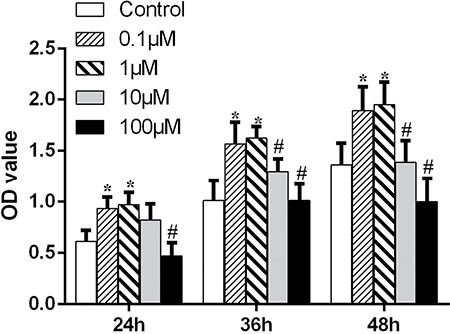
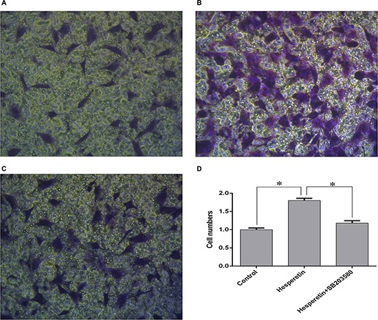
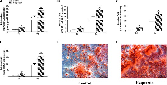
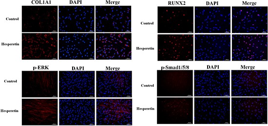
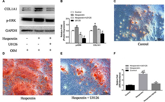
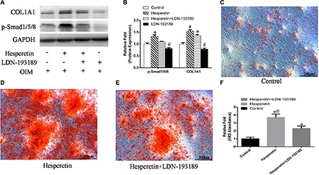
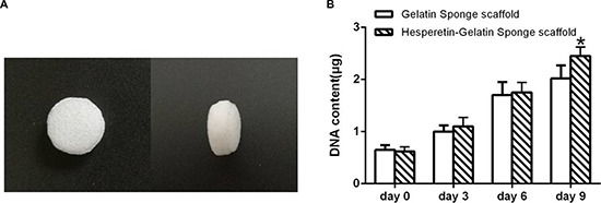
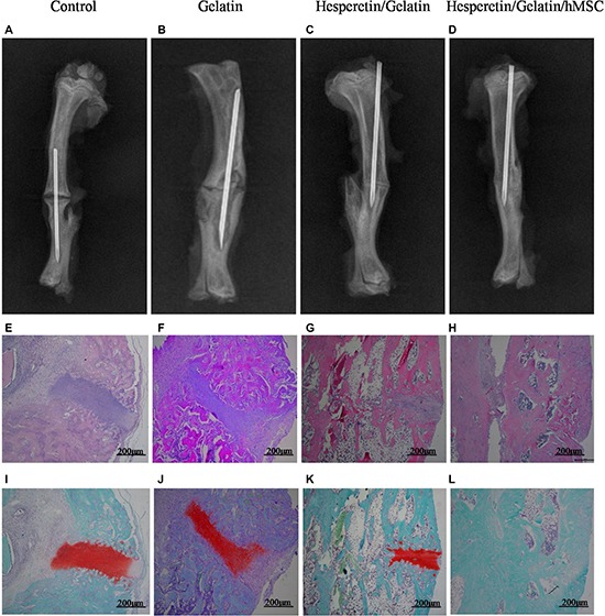
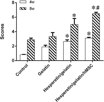
Hesperetin has been suggested to be involved in bone strength. We aimed to investigate the effects of hesperetin on the osteogenic differentiation of human mesenchymal stem cells and its related mechanisms. We showed that hesperetin promoted osteogenic differentiation of human mesenchymal stem cells in vitro. It potentially exerts its effects via the ERK and Smad signaling pathways. Using a rat osteotomy model, we showed that human mesenchymal stem cells combined with a hesperetin/gelatin sponge scaffold resulted in accelerated fracture healing in vivo. Due to the low cost of hesperetin, it could be used as a growth factor for bone tissue engineering or surgical fracture treatment.
Keywords: bone regeneration; gelatin sponge scaffold; hesperetin; mesenchymal stem cells; osteogenesis.
Conflict of interest statement
The authors declare no conflicts of interest.
Figures
Similar articles
-
Osteogenic Potential of Mouse Periosteum-Derived Cells Sorted for CD90 In Vitro and In Vivo.Stem Cells Transl Med. 2016 Feb;5(2):227-34. doi: 10.5966/sctm.2015-0013. Epub 2015 Dec 30. Stem Cells Transl Med. 2016. PMID: 26718647 Free PMC article.
-
Characterization of human ethmoid sinus mucosa derived mesenchymal stem cells (hESMSCs) and the application of hESMSCs cell sheets in bone regeneration.Biomaterials. 2015 Oct;66:67-82. doi: 10.1016/j.biomaterials.2015.07.013. Epub 2015 Jul 14. Biomaterials. 2015. PMID: 26196534
-
Administration of tauroursodeoxycholic acid enhances osteogenic differentiation of bone marrow-derived mesenchymal stem cells and bone regeneration.Bone. 2016 Feb;83:73-81. doi: 10.1016/j.bone.2015.10.011. Epub 2015 Oct 21. Bone. 2016. PMID: 26499839
-
Extracellular Signals for Guiding Mesenchymal Stem Cells Osteogenic Fate.Curr Stem Cell Res Ther. 2017;12(2):139-144. doi: 10.2174/1574888X10666151026114411. Curr Stem Cell Res Ther. 2017. PMID: 26496887 Review.
-
Nanomaterials-based Cell Osteogenic Differentiation and Bone Regeneration.Curr Stem Cell Res Ther. 2021;16(1):36-47. doi: 10.2174/1574888X15666200521083834. Curr Stem Cell Res Ther. 2021. PMID: 32436831 Review.
Cited by
-
Mitigation of BMP-induced inflammation in craniofacial bone regeneration and improvement of bone parameters by dietary hesperidin.Sci Rep. 2024 Jan 31;14(1):2602. doi: 10.1038/s41598-024-52566-7. Sci Rep. 2024. PMID: 38297106 Free PMC article.
-
Traditional Chinese medicine promotes bone regeneration in bone tissue engineering.Chin Med. 2022 Jul 20;17(1):86. doi: 10.1186/s13020-022-00640-5. Chin Med. 2022. PMID: 35858928 Free PMC article. Review.
-
Effect of Flavonoid Supplementation on Alveolar Bone Healing-A Randomized Pilot Trial.Dent J (Basel). 2020 Aug 4;8(3):86. doi: 10.3390/dj8030086. Dent J (Basel). 2020. PMID: 32759635 Free PMC article.
-
Polymeric and Composite Carriers of Protein and Non-Protein Biomolecules for Application in Bone Tissue Engineering.Materials (Basel). 2023 Mar 10;16(6):2235. doi: 10.3390/ma16062235. Materials (Basel). 2023. PMID: 36984115 Free PMC article. Review.
-
Antioxidant, Anti-Inflammatory, Anti-Diabetic, and Pro-Osteogenic Activities of Polyphenols for the Treatment of Two Different Chronic Diseases: Type 2 Diabetes Mellitus and Osteoporosis.Biomolecules. 2024 Jul 11;14(7):836. doi: 10.3390/biom14070836. Biomolecules. 2024. PMID: 39062550 Free PMC article. Review.
References
-
- Phieffer LS, Goulet JA. Delayed unions of the tibia. J Bone Joint Surg Am. 2006;88:206–216. - PubMed
-
- Velazco A, Whitesides TE, Jr, Fleming LL. Open fractures of the tibia treated with the Lottes nail. J Bone Joint Surg Am. 1983;65:879–885. - PubMed
-
- Clancey GJ, Hansen ST., Jr Open fractures of the tibia: a review of one hundred and two cases. J Bone Joint Surg Am. 1978;60:118–122. - PubMed
-
- Bell A, Templeman D, Weinlein JC. Nonunion of the Femur and Tibia: An Update. Orthop Clin North Am. 2016;47:365–375. - PubMed
-
- Govender S, Csimma C, Genant HK, Valentin-Opran A, Amit Y, Arbel R, Aro H, Atar D, Bishay M, Borner MG, Chiron P, Choong P, Cinats J, et al. Recombinant human bone morphogenetic protein-2 for treatment of open tibial fractures: a prospective, controlled, randomized study of four hundred and fifty patients. J Bone Joint Surg Am. 2002;84-A:2123–2134. - PubMed
MeSH terms
Substances
LinkOut - more resources
Full Text Sources
Other Literature Sources
Miscellaneous
