. 2017 Mar 28;8(13):21719-21732.
doi: 10.18632/oncotarget.15559.

miR-503-5p confers drug resistance by targeting PUMA in colorectal carcinoma

Affiliations

miR-503-5p confers drug resistance by targeting PUMA in colorectal carcinoma

Ke Xu et al. Oncotarget. .

Abstract

The development of multidrug-resistance (MDR) is a major contributor to death in colorectal carcinoma (CRC). Here, we investigated the possible role of microRNA (miR)-503-5p in drug resistant CRC cells. Unbiased microRNA array screening revealed that miR-503-5p is up-regulated in two oxaliplatin (OXA)-resistant CRC cell lines. Overexpression of miR-503-5p conferred resistance to OXA-induced apoptosis and inhibition of tumor growth in vitro and in vivo through down-regulation of PUMA expression. miR-503-5p knockdown sensitized chemoresistant CRC cells to OXA. Our studies indicated that p53 suppresses miR-503-5p expression and that deletion of p53 upregulates miR-503-5p expression. Inhibition of miR-503-5p in p53 null cells increased their sensitivity to OXA treatment. Importantly, analysis of patient samples showed that expression of miR-503-5p negatively correlates with PUMA in CRC. These results indicate that a p53/miR-503-5p/PUMA signaling axis regulates the CRC response to chemotherapy, and suggest that miR-503-5p plays an important role in the development of MDR in CRC by modulating PUMA expression.

Keywords: PUMA; colorectal carcinoma; miR-503-5p; multidrug-resistance; p53.


Conflict of interest statement

CONFLICTS OF INTEREST

The authors declare no competing financial interest.

Figures
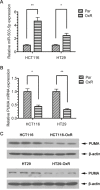
Figure 1
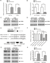
Figure 1. Expression of miR-503-5p and PUMA in human CRC cell lines
A. qPCR validation of miR-503-5p expression levels significantly increased in oxaliplatin-resistant (OXR) CRC cells. B. qPCR validation of PUMA mRNA expression levels significantly decreased in oxaliplatin-resistant (OXR) CRC cells. C. Western blot validation of PUMA protein expression levels significantly decreased in oxaliplatin-resistant (OXR) CRC cells. Abundance of miR-503-5p and PUMA was normalized to U6 RNA, GAPDH and β-actin, respectively. Results are representative of three experiments.
Figure 2
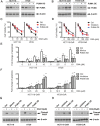
Figure 2. PUMA is a direct target of miR-503-5p
A. MiR-503-5p expression level was significantly increased after transient transfection of miR-503-5p mimic in CRC parental cells as measured by qPCR. B. MiR-503-5p expression level was significantly decreased after transient transfection of miR-503-5p inhibitor in in CRC oxaliplatin-resistant cells as measured by qPCR C. PUMA protein expression levels are regulated directly by miR-503-5p, as reflected by decreased PUMA expression in CRC-Par cells after transient transfection of miR-503-5p mimic D. and increased PUMA expression in CRC-OxR cells after miR-503-5p inhibitor transfection. E. The wild-type and mutant variant of the putative miR-503-5p target sequences of the PUMA gene. TargetScan predicts one binding sites in the PUMA 3′UTR. F. Two copies of the wild-type and mutant miR-503-5p target sequences were fused with a luciferase reporter and transfected into control oligonucleotide and 40 nM miR-503-5p mimics infected HCT116-OxR cells. MiR-503-5p significantly suppressed the luciferase activity of the wild-type PUMA 3′UTR. G. Bcl-2, Mcl-1 and Bax protein levels were assessed 48 h after transfection of control oligonucleotide and MiR-503-5p inhibitors (40 nM) HCT116-OxR cells, which were respectively detected by Western blotting. H. The percentage of caspases-3 activity was assessed in HCT116-OxR cells as described above. **p<0.01. Results are representative of three experiments.
Figure 3
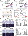
Figure 3. Modulation of miR-503-5p expression altered the sensitivity of CRC cells to oxaliplatin in vitro
A. Expression of PUMA was knocked down by shRNAs in CRC-Par cells. B. Expression of PUMA was overexpression by Plasmids in CRC-OxR cells. C. Overexpression of miR-503-5p induced by mimics transfection significantly decreased the growth-inhibitory effect of oxaliplatin in CRC-Par cells by MTT assay; E. decreased CRC-Par cells apoptosis level to oxaliplatin by DNA fragmentation assays; G. and decreased cleaved PARP level by western blot analysis, same effect as PUMA knock down (PUMA KD). D. Down-regulation of miR-503-5p induced by inhibitors transfection significantly slowed down CRC-OxR cells growth under oxaliplatin treatment by MTT assay; F. increased CRC-OxR cells apoptosis level to oxaliplatin by DNA fragmentation assays; H. and increased cleaved PARP level by western blot analysis, same effect as PUMA overexpression (PUMA OE). *p<0.05, **p<0.01. Results are representative of three experiments.
Figure 4
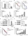
Figure 4. Modulation of miR-503-5p expression altered the sensitivity of CRC cells to oxaliplatin in vivo
Overexpression of miR-503-5p reduced the effectiveness of OXA in the inhibition of tumor growth in vivo. A. Xenograft tumor growth curves, C. (up panel) pictures of tumors taken on the same scale D. and tumor weights. Down-regulation of miR-503-5p increased the effectiveness of OXA in the inhibition of tumor growth in vivo. B. Xenograft tumor growth curves, C. (down panel) pictures of tumors taken on the same scale E. and tumor weights. F. Overexpression of miR-503-5p significantly decreased the growth-inhibitory effect of oxaliplatin in CRC-Par cells by Ki67 level, and decreased CRC-Par cells apoptosis level to oxaliplatin by TUNEL level in vivo. G. Down-regulation of miR-503-5p significantly slowed down CRC-OxR cells growth under oxaliplatin treatment by Ki67 level, and increased CRC-OxR cells apoptosis level to oxaliplatin by TUNEL level in vivo. The images are representative of multiple fields of tumor sections from each group. Percentage of positive Ki67 and TUNEL staining cells were determined as described in Materials and Methods. The data are presented as the mean ± SD. *p < 0.05, **p<0.01, *** p < 0.001.
Figure 5
Figure 5. p53 suppresses miR-503-5p expression
A. miR-503-5p expression was higher in p53 knock out (KO) than wild type (WT) HCT116 cells, and decreased in the p53 overexpression (OE) HCT116-OxR cells compared to control plasmid, by qPCR assays. WT and p53 KO HCT116 cells, Ctrl and p53 OE HCT116-OxR cells were treated with different concentrations of OXA for 48 hrs. B. MTT assays and C. DNA fragmentation assays showed that HCT116 p53 KO cells were more resistant to OXA-induced apoptosis than WT cells, and p53 OE HCT116-OxR cells were more sensitive to OXA-induced apoptosis than Ctrl cells. D. p53 was ectopically expressed in HCT116 p53 KO cells. Western blot analysis showed restored expression of p53 and PUMA expression (left panel), qPCR assays showed that restored p53 expression suppressed miR-503-5p expression (right panel).E. The miR-503-5p inhibitor was transfected into HCT116 p53 KO cells. As a result, miR-503-5p expression was suppressed as determined by qPCR assays (left panel) and PUMA expression was increased as examined by western blot assays (right panel). Cells were treated with different concentrations of OXA for 48 hrs. F. MTT assays and G. DNA fragmentation assays showed that restored expression of p53 and miR-503-5p inhibitor re-sensitized p53 KO cells to OXA-induced apoptosis. The data are presented as the mean ± SD of triplicate experiments. *p < 0.05, **p<0.01, *** p < 0.001.
Figure 6
Figure 6. Expression of miR-503-5p and PUMA in human CRC tissue sample
Relative expression levels of A. miR-503-5p and B. PUMA mRNA were detected in non-tumorous colon (NC) tissues and CRC tissues (n=29) via qRT-PCR. Abundance of miR-503-5p and PUMA was normalized to U6 RNA and GAPDH, respectively. C. Expression levels of miR-503-5p and PUMA mRNA are inversely correlated among all the tissue samples (n = 29) as indicated by two-tailed Pearson's correlation analysis, r = −0.58; p < 0.01. D. miR-503-5p expression levels were inversely correlated with PUMA mRNA expression in CRC tissues. E & F. PUMA protein level were detected in normal colon tissues (NC) and CRC tissues (n=29) via Western blot analysis. Abundance PUMA was normalized to β-actin. G. Expression levels of miR-503-5p and PUMA protein level are inversely correlated among all the tissue samples (n = 29) as indicated by two-tailed Pearson's correlation analysis, r = −0.81; p < 0.01. H. miR-503-5p expression levels were inversely correlated with PUMA protein level expression in CRC tissues. The data are presented as the mean ± SD. *p < 0.05, **p<0.01, *** p < 0.001.

Similar articles

Cited by

References

    1. Obuch JC, Ahnen DJ. Colorectal Cancer: Genetics is Changing Everything. Gastroenterology clinics of North America. 2016;45:459–476. - PubMed
    1. Meyerhardt JA, Mayer RJ. Systemic therapy for colorectal cancer. The New England journal of medicine. 2005;352:476–487. - PubMed
    1. Wang D, Lippard SJ. Cellular processing of platinum anticancer drugs. Nature reviews Drug discovery. 2005;4:307–320. - PubMed
    1. Hector S, Bolanowska-Higdon W, Zdanowicz J, Hitt S, In Pendyala L. vitro studies on the mechanisms of oxaliplatin resistance. Cancer chemotherapy and pharmacology. 2001;48:398–406. - PubMed
    1. Landriscina M, Maddalena F, Laudiero G, Esposito F. Adaptation to oxidative stress, chemoresistance, and cell survival. Antioxidants & redox signaling. 2009;11:2701–2716. - PubMed

MeSH terms