. 2017 Mar 28;8(13):21778-21793.
doi: 10.18632/oncotarget.15583.

Vitamin D and VDR in cancer cachexia and muscle regeneration

Affiliations

Vitamin D and VDR in cancer cachexia and muscle regeneration

Andrea Camperi et al. Oncotarget. .

Abstract

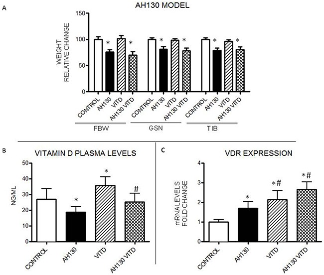
Low circulating levels of vitamin D were associated with decreased muscle strength and physical performance. Along this line, the present study was aimed to investigate: i) the therapeutic potential of vitamin D in cancer-induced muscle wasting; ii) the mechanisms by which vitamin D affects muscle phenotype in tumor-bearing animals.Rats bearing the AH130 hepatoma showed decreased circulating vitamin D compared to control rats, while muscle vitamin D receptor (VDR) mRNA was up-regulated. Both circulating vitamin D and muscle VDR expression increased after vitamin D administration, without exerting appreciable effects on body weight and muscle mass.The effects of vitamin D on muscle cells were studied in C2C12 myocytes. Vitamin D-treated myoblasts did not differentiate properly, fusing only partially and forming multinucleated structures with aberrant shape and low myosin heavy chain content. Vitamin D treatment resulted in VDR overexpression and myogenin down-regulation. Silencing VDR expression in C2C12 cultures abrogated the inhibition of differentiation exerted by vitamin D treatment.These data suggest that VDR overexpression in tumor-bearing animals contributes to muscle wasting by impairing muscle regenerative program. In this regard, attention should be paid when considering vitamin D supplementation to patients affected by chronic pathologies where muscle regeneration may be involved.

Keywords: circulating vitamin D; muscle wasting; myogenin; regeneration; vitamin D receptor.


Conflict of interest statement

CONFLICTS OF INTEREST

The authors have no conflict of interest to declare.

Figures

Figure 1
Figure 1. Effects of VitD3 administration to rats bearing the AH130 hepatoma
A. Body weight changes, gastrocnemius and tibialis muscle weight in controls and tumor-bearing rats either untreated or receiving VitD3. B. VitD plasma levels in untreated and VitD3-treated rats. C. VDR mRNA expression levels in the tibialis muscle of untreated and VitD3-treated animals. Data (means±SEM) are expressed as fold change (control rats: n=6, tumor-bearing rats: n=8). Significance of the differences: *p< 0.05 vs control; #p< 0.05 vs AH130.
Figure 2
Figure 2. VitD levels and VDR expression in mice bearing the LLC or the C26 tumor
A, D. Final body weight (FBW), gastrocnemius (GSN) and tibialis (TIB) mass in controls (n=5) and tumor-bearing mice (n=7). B, E. Circulating VitD and VDR protein expression levels in GSN muscle. C, F. Representative WB analysis of VDR expression in the gastrocnemius (anti-VDR antibody, clone D6, Santa Cruz Biotechnology). Data are means±SD. Significance of the differences: *p< 0.05 vs C.
Figure 3
Figure 3. Effects of VitD on C2C12 myoblast differentiation
A. Immunostaining of control or VitD-treated C2C12 cells after 4 days of differentiation (Red: MyHC, Blue: nuclei). B. Expression levels of VDR (black line) and myogenin (grey line) during C2C12 myoblast differentiation in the absence of VitD treatment, as measured by western blotting analysis (Santa Cruz Biotechnology: anti-VDR antibody, clone D6; anti-myogenin antibody, clone F5D). C. Protein expression levels of some myogenic regulatory factors in control or VitD-stimulated C2C12 cells at day 2 and 4 of differentiation (Santa Cruz Biotechnology: anti-myogenin antibody, clone F5D, anti-MyoD antibody, clone M318; Developmental Studies Hybridoma Bank, University of Iowa: anti-Pax7 antibody). Data (means±SD) are expressed as % of controls. Significance of the differences: ***p<0.001, **p<0.01, *p<0.05 vs C, 3 independent experiments.
Figure 4
Figure 4. Effects of VitD on VDR and myogenin expression on C2C12 differentiating myoblasts and on myotubes
A. Protein expression levels of VDR and myogenin in control or VitD-stimulated C2C12 cells at day 4 of differentiation (Santa Cruz Biotechnology: anti-VDR antibody, clone D6, anti-myogenin antibody, clone F5D); B. Average myotube diameter and average number of nuclei/myotube in C2C12 cultures treated with control medium or with medium containing: 100ng/ml IL6, 10nM or 100nM VitD (bars show the average of three independent experiments, n=100 for each condition); C. Western blotting analysis of representative samples showing VDR expression (Santa Cruz Biotechnology: anti-VDR antibody, clone D6). Data are means ± SD. Significance of the differences: *p< 0.05 vs C, 3 independent experiments.
Figure 5
Figure 5. VDR is recruited at regulatory regions of the myogenin gene after VitD exposition
A. Putative VDR binding sites in the promoter region of the Myogenin gene (6.5 Kbp), as predicted by LASAGNA-Search web tool, and schematic representation of the Myogenin promoter region showing putative binding sites for VDR; B. ChIP assay (representative pattern) was performed with chromatin extracted from C2C12 myoblasts treated or untreated with VitD, using normal rabbit IgG and an anti-VDR antibody. β-globin and cytochrome c promoter regions were amplified as negative and positive controls, respectively.
Figure 6
Figure 6. Effects of VitD treatment on VDR-silenced C2C12 cells
A. Immunostaining of C2C12 cells after 4 days of differentiation (Red: MyHC, Blue: nuclei). B. Protein expression levels of VDR, MyHC and myogenin at day 4 of differentiation. Data (means±SD) are expressed as relative change. C. number of nuclei/myofiber. D. representative western blotting pattern of MyHC, myogenin and VDR expression (Santa Cruz Biotechnology: anti-VDR antibody, clone D6, anti-myogenin antibody, clone F5D; Sigma: anti MyHC antibody, clone MY32). Significance of the differences: *p<0.05 vs ShC, 2 independent experiments.
Figure 7
Figure 7. Effects of VitD administration during muscle regeneration
A. Histological pattern of regenerating and non regenerating muscles in VitD3-treated (n = 6) and untreated mice (n = 6). For experimental details see Materials and Methods. B. VDR expression in regenerating and non regenerating muscles in the presence or in the absence of VitD3 treatment (Santa Cruz Biotechnology: anti-VDR antibody, clone D6). Data (means±SD) are expressed as relative change. Significance of the differences: *p< 0.05 vs C. C. Average myofiber CSA of controls (white bars) or regenerating (black bars) muscles 15 days after BaCl2 injection; 400 myofibers (regenerating or not)/condition have been counted. D. Representative samples showing VDR, Pax7 and myogenin expression (Santa Cruz Biotechnology: anti-VDR antibody, clone D6, anti-myogenin antibody, clone F5D; Developmental Studies Hybridoma Bank, University of Iowa: anti-Pax7 antibody).

References

    1. Muscaritoli M, Molfino A, Lucia S, Rossi Fanelli F. Cachexia: a preventable comorbidity of cancer A T.A.R.G.E.T. approach. Crit Rev Oncol Hematol. 2015;94:251–259. - PubMed
    1. Argiles JM, Busquets S, Stemmler B, Lopez-Soriano FJ. Cancer cachexia: understanding the molecular basis. Nat Rev Cancer. 2014;14:754–762. - PubMed
    1. Suzuki H, Asakawa A, Amitani H, Nakamura N, Inui A. Cancer cachexia--pathophysiology and management. J Gastroenterol. 2013;48:574–594. - PMC - PubMed
    1. Baracos VE, DeVivo C, Hoyle DH, Goldberg AL. Activation of the ATP-ubiquitin-proteasome pathway in skeletal muscle of cachectic rats bearing a hepatoma. Am J Physiol. 1995;268:E996–1006. - PubMed
    1. He WA, Berardi E, Cardillo VM, Acharyya S, Aulino P, Thomas-Ahner J, Wang J, Bloomston M, Muscarella P, Nau P, Shah N, Butchbach ME, Ladner K, et al. NF-kappaB-mediated Pax7 dysregulation in the muscle microenvironment promotes cancer cachexia. J Clin Invest. 2013;123:4821–4835. - PMC - PubMed

MeSH terms