. 2017 May 9;8(19):31785-31801.
doi: 10.18632/oncotarget.15991.

The novel long non-coding RNA TALNEC2, regulates tumor cell growth and the stemness and radiation response of glioma stem cells

Affiliations

The novel long non-coding RNA TALNEC2, regulates tumor cell growth and the stemness and radiation response of glioma stem cells

Shlomit Brodie et al. Oncotarget. .

Erratum in

Abstract

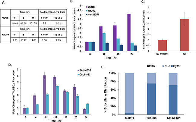
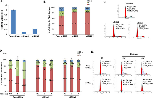
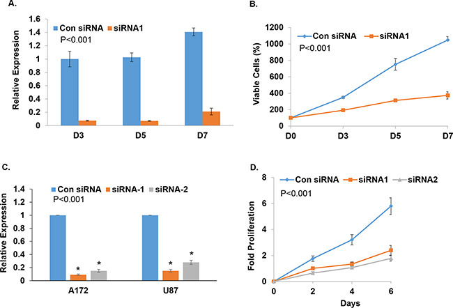
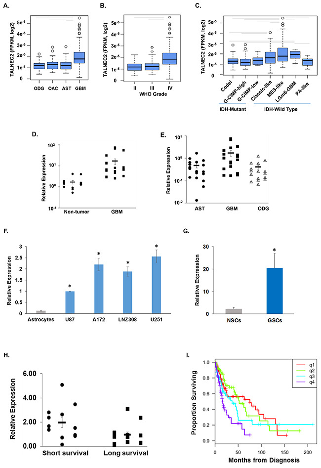
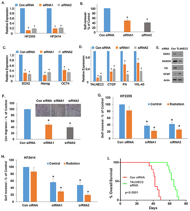
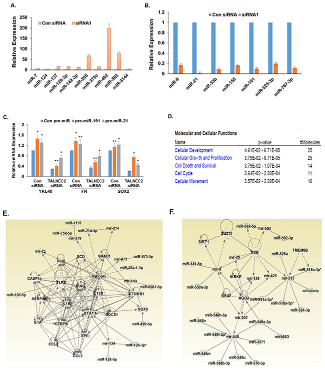
Despite advances in novel therapeutic approaches for the treatment of glioblastoma (GBM), the median survival of 12-14 months has not changed significantly. Therefore, there is an imperative need to identify molecular mechanisms that play a role in patient survival. Here, we analyzed the expression and functions of a novel lncRNA, TALNEC2 that was identified using RNA seq of E2F1-regulated lncRNAs. TALNEC2 was localized to the cytosol and its expression was E2F1-regulated and cell-cycle dependent. TALNEC2 was highly expressed in GBM with poor prognosis, in GBM specimens derived from short-term survivors and in glioma cells and glioma stem cells (GSCs). Silencing of TALNEC2 inhibited cell proliferation and arrested the cells in the G1\S phase of the cell cycle in various cancer cell lines. In addition, silencing of TALNEC2 decreased the self-renewal and mesenchymal transformation of GSCs, increased sensitivity of these cells to radiation and prolonged survival of mice bearing GSC-derived xenografts. Using miRNA array analysis, we identified specific miRNAs that were altered in the silenced cells that were associated with cell-cycle progression, proliferation and mesenchymal transformation. Two of the downregulated miRNAs, miR-21 and miR-191, mediated some of TALNEC2 effects on the stemness and mesenchymal transformation of GSCs. In conclusion, we identified a novel E2F1-regulated lncRNA that is highly expressed in GBM and in tumors from patients of short-term survival. The expression of TALNEC2 is associated with the increased tumorigenic potential of GSCs and their resistance to radiation. We conclude that TALNEC2 is an attractive therapeutic target for the treatment of GBM.

Keywords: TALNEC2; glioblastoma; glioma stem cells; long non-cording RNAs; mesenchymal transformation.


Conflict of interest statement

CONFLICTS OF INTEREST

The authors declare no conflicts of interest.

Figures

Figure 1
Figure 1. Characterization of TALNEC2 expression and subcellular localization
RNA Seq. results for TALNEC2 are shown as fold of induction after addition of OHT for 8 and 16 h compared to untreated cells (A). U2OS and H1299 cells expressing ER-E2F1 and U2OS cells expressing mutated ER-E2F1 were treated with OHT for the indicated time points (in hours). RNA was extracted and TALNEC2 RNA levels were determined by real-time PCR and normalized to GAPDH levels. Real-time PCR experiments were performed in triplicates (B). WI38 cells were infected with a retrovirus vector expressing either wild-type E7 (E7) or an RB-binding–deficient mutant of E7 (E7-mutant). RNA was extracted and TALNEC2 RNA levels were determined by Real-time PCR and normalized to GAPDH levels. Real-time PCR experiments were performed in duplicates (C). WI38 cells were growth arrested by serum deprivation (48 hours in 0.1% serum) and then allowed to resume growth by serum addition (to a final concentration of 15%) for the indicated times. RNA was extracted and levels of CCNE1 and TALNEC2 RNA levels were determined by Real-time PCR and normalized to GAPDH levels. CCNE1 is a known E2F-regulated gene serving as a positive control for cell cycle-dependent gene expression. Real-time PCR experiments were performed in triplicates (D). RNA was extracted from the nuclear and cytoplasmic fractions of the U2OS cells as described in the methods and the levels of nuclear control transcript (MALAT1), cytoplasmic control transcript (GAPDH or tubulin), and TALNEC2 were determined by RT-PCR in the nuclear and cytoplasmic fractions. The bar graphs present the percentage of nuclear and cytoplasmic RNAs out of the total cell RNA. Real-time PCR experiments were performed in triplicate (E). The results are representative of three different experiments that gave similar results.
Figure 2
Figure 2. Silencing of TALNEC2 arrests cells in the G1 phase of the cell cycle
MCF-7 cells were transfected with either a nonspecific siRNA (Con siRNA, 100nM ) or siRNAs directed against TALNEC2 (siRNA1 (25nM), siRNA2 (100nM)). Cells were harvested 72 hours post transfection (A, B, C). RNA was extracted and TALNEC2 RNA levels were determined by real-time PCR and normalized to GAPDH levels. Real-time PCR experiments were performed in triplicates (A). Cells were analyzed by FACS using propidium-iodide (PI) protocol. Percentages of cells in G1, S, and G2/M cell-cycle phases are shown (B, C). The results are either representative (C) or average of four independent experiments (B). *P<0.05, **P<0.01 (two-tailed students t-test). MCF-7 or H1299 cells were transfected with either a nonspecific siRNA (Con siRNA, 100nM) or siRNAs directed against TALNEC2 (siRNA1 (25nM), siRNA2 (100nM). Next, cells were incubated with hydroxyurea (HU, 2mM) for 18 h. 72 h post transfection cells were harvested or allowed to resume growth by fresh media wash and growth in the fresh media for times indicated. Cells were analyzed by FACS using propidium-iodide (PI) protocol (D, E). Percentages of cells in G1, S, and G2-M cell-cycle phases are depicted. (D, E). Average FACS analysis for MCF-7 cells is for four independent experiments (D). *P<0.05, **P<0.01 (two-tailed students t-test). For H1299 cells, the results are representative of three independent experiments (E).
Figure 3
Figure 3. Silencing of TALNEC2 inhibits cell proliferation
MCF-7 cells were transfected with either a nonspecific siRNA (Con siRNA, 25nM ) or siRNA directed against TALNEC2 (siRNA1, 25nM). Cells were harvested at the indicated days post transfection (A, B). RNA was extracted and TALNEC2 RNA levels were determined by Real-time PCR and normalized to GAPDH levels. Real-time PCR experiments were performed in triplicates (A). Cell proliferation was measured by MTT assay (B). U87 and A172 glioma cells were transfected with either a nonspecific siRNA (Con siRNA) or siRNAs directed against TALNEC2 (siRNA1, siRNA2). Levels of TALNEC2 were determined after 3 days using RT-PCR (C) and cell proliferation was determined for the U87 cells at the indicated time points using MTT (D). The results are representative of three different experiments that gave similar results.*P<0.001 **P< 0.01.
Figure 4
Figure 4. Expression of TALNEC2 in GBM, glioma cell lines and GSCs
Expression of TALNEC2 in glioma tissues was determined from RNA-sequencing data from The Cancer Genome Atlas project. The distribution of expression is shown by boxplots for histology (A), WHO grade (B), and supervised IDH-methylation classes (C). Average expression was compared by ANOVA (P<0.0001) with follow-up t-tests (P<0.05, grey bars). Total RNA was extracted from normal brains (Non-tumor), astrocytoma (AST), oligodendroglioma (ODG) and GBM specimens and the expression of TALNEC2 was determined using real-time PCR (D, E). Results are normalized relative to the levels of S12 mRNA and are presented relative to a reference sample as dot-plots around the estimate of the mean and SEM bars. GBM has higher TALNEC2 expression, on average, than non-tumor brain, AST, or ODG (P<0.01). The mean expression of TALNEC2, measured using real-time PCR, in glioma cell lines relative to human astrocytes (F) is shown by barplots with SEM bars (student test P<0.001 indicated by *). Twelve different primary GSCs, generated from GBM specimens, show increased mean expression of TALNEC2 (by RT-PCR), relative to human NSCs (G, P<0.001). GBM specimens obtained from sort-term survivors (<9 months, n=13) have higher mean TALNEC2 expression compared to long-term survivors (>36 months, n=12; P< 0.01, H). Kaplan-Meier survival estimates of overall survival are plotted for patients grouped by TALNEC2 expression quartiles (q1=lowest expression; log-rank p= 0.000064, I). The results are presented as the mean values ± SD. Data were analyzed using analysis of variance or a Student's t-test.
Figure 5
Figure 5. TALNEC2 regulates GSC stemness, mesenchymal transformation and response to radiation
GSCs transfected with a control or TALNEC2 siRNAs were analyzed for the expression of TALNEC2 (A). The HF2355 GSCs were then plated at 100 cells/well in 96-well plates and the number of neurospheres per well was quantified after 14 days (B). P < 0.0001. The expression of the stemness markers, NANOG, SOX2 and OCT4 (C), and the mesenchymal markers, CTGF, fibronectin and YKL40 were determined using qPCR (D) or western blot assay (E). The results are representative of three different experiments that gave similar results. Cell migration was assayed using a transwell chamber containing polycarbonate membrane inserts of 8-μm pore size (Corning, Costar) with fibronectin coating as described in the methods. The HF2355 GSCs transfected with a con siRNA or silenced for TALNEC2 were analyzed in this assay. The results are shown are the mean ± SE of triplicate experiments (F) (P<0.01). The HF2355 (G) and HF2414 (H) GSCs transfected with control or TALNEC2 siRNAs were irradiated (3 Gy) and self-renewal was analyzed after 2 weeks (G, H). Kaplan-Meier survival curves for mice transplanted with the HF2587 GSCs transfected with a control siRNA or TALNEC2 siRNA1 (n = 11) were determined by both log-rank (Mantel-Cox) test and Gehan-Breslow-Wilcoxon test (I).
Figure 6
Figure 6. Alteration in miRNA expression in glioma cells silenced for TALNEC2
U87 cells were silenced for TALNEC2 using siRNA1 and siRNA2 for 2 days and the cells were analyzed for miRNA expression miRNome microRNA Profilers QuantiMir™. Validation for specific miRNAs that were altered in the TALNEC2 silenced U87 cells was performed for the HF2355 GSCs using RT-PCR. P<0.001 (A, B). The role of miR-21 and miR-191 in TALNEC2 effects was examined by the transduction of TALNEC2 silenced HF2355 GSCS with lentivirus vectors expressing pre-miR-21 and pre-mir-191. The expression of stemness and mesenchymal markers was determined using RT-PCR (C). Ingenuity functional clustering analysis (D) and generation of Ingenuity networks (E, F) were performed for the altered miRNAs in the TALNEC2-silenced glioma cells.

Similar articles

Cited by

References

    1. Iyer MK, Niknafs YS, Malik R, Singhal U, Sahu A, Hosono Y, Barrette TR, Prensner JR, Evans JR, Zhao S, Poliakov A, Cao X, Dhanasekaran SM, et al. The landscape of long noncoding RNAs in the human transcriptome. Nature genetics. 2015;47:199–208. - PMC - PubMed
    1. Brunner AL, Beck AH, Edris B, Sweeney RT, Zhu SX, Li R, Montgomery K, Varma S, Gilks T, Guo X, Foley JW, Witten DM, Giacomini CP, et al. Transcriptional profiling of long non-coding RNAs and novel transcribed regions across a diverse panel of archived human cancers. Genome biology. 2012;13:R75. - PMC - PubMed
    1. Sahu A, Singhal U, Chinnaiyan AM. Long noncoding RNAs in cancer: from function to translation. Trends Cancer. 2015;1:93–109. - PMC - PubMed
    1. Huarte M. The emerging role of lncRNAs in cancer. Nat Med. 2015;21:1253–1261. - PubMed
    1. Schmitt AM, Chang HY. Long Noncoding RNAs in Cancer Pathways. Cancer Cell. 2016;29:452–463. - PMC - PubMed

MeSH terms