. 2017 May 23;8(21):34082-34098.
doi: 10.18632/oncotarget.16133.

Honokiol, an activator of Sirtuin-3 (SIRT3) preserves mitochondria and protects the heart from doxorubicin-induced cardiomyopathy in mice

Affiliations

Honokiol, an activator of Sirtuin-3 (SIRT3) preserves mitochondria and protects the heart from doxorubicin-induced cardiomyopathy in mice

Vinodkumar B Pillai et al. Oncotarget. .

Abstract

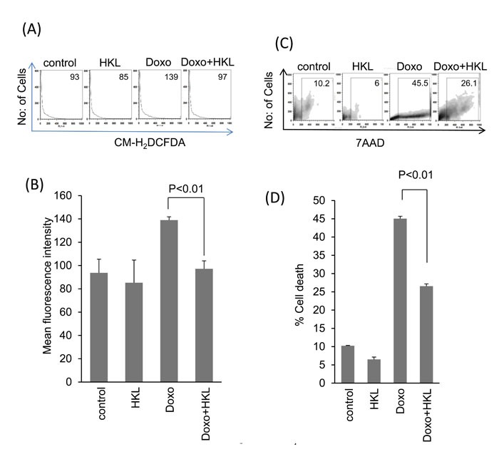
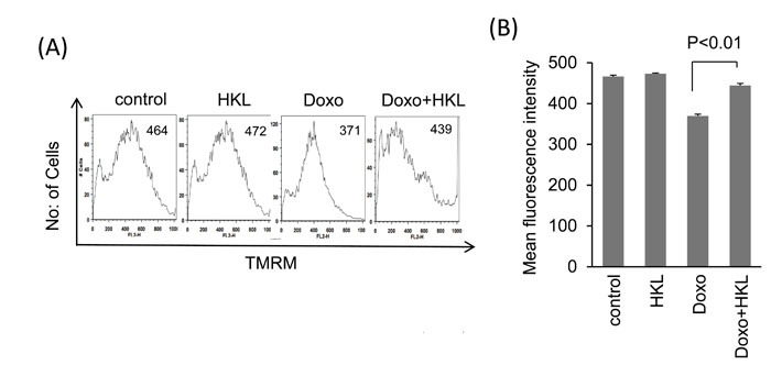
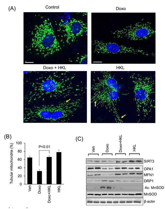
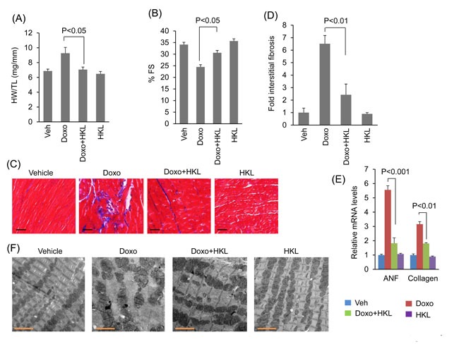
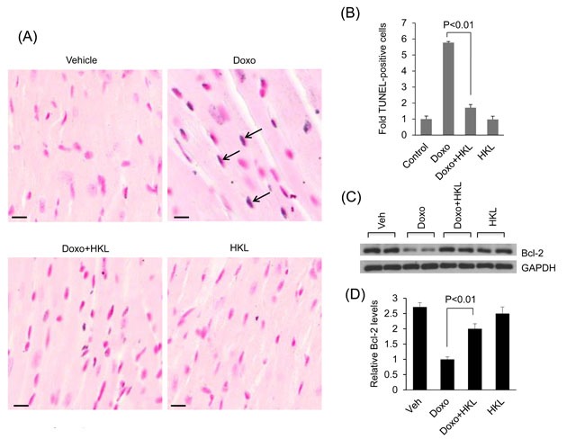
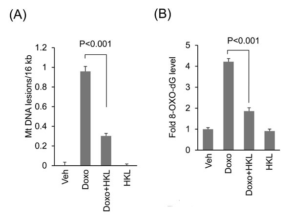
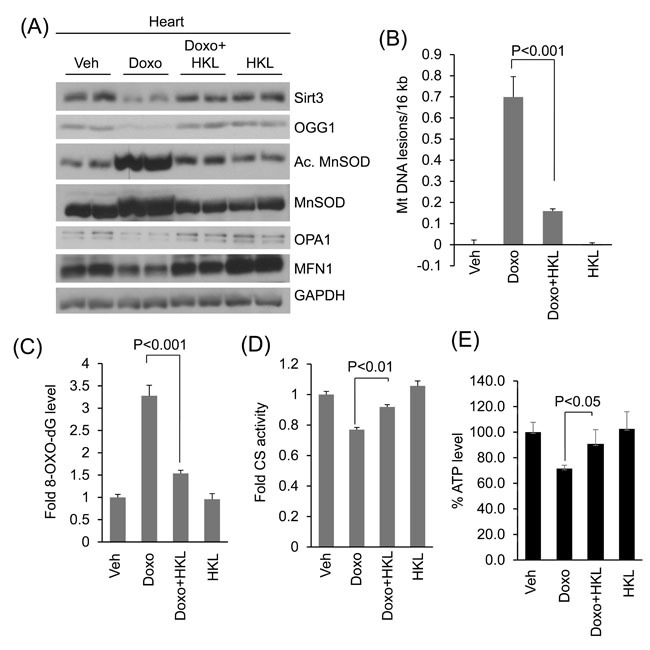
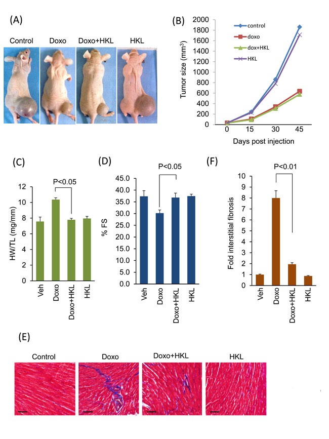
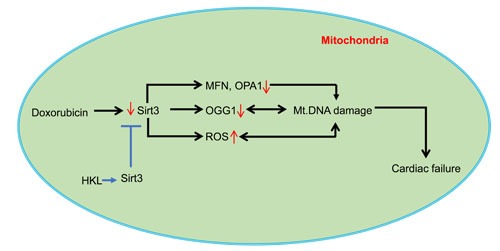
Doxorubicin is the chemotherapeutic drug of choice for a wide variety of cancers, and cardiotoxicity is one of the major side effects of doxorubicin treatment. One of the main cellular targets of doxorubicin in the heart is mitochondria. Mitochondrial sirtuin, SIRT3 has been shown to protect against doxorubicin-induced cardiotoxicity. We have recently identified honokiol (HKL) as an activator of SIRT3, which protects the heart from developing pressure overload hypertrophy. Here, we show that HKL-mediated activation of SIRT3 also protects the heart from doxorubicin-induced cardiac damage without compromising the tumor killing potential of doxorubicin. Doxorubicin-induced cardiotoxicity is associated with increased ROS production and consequent fragmentation of mitochondria and cell death. HKL-mediated activation of SIRT3 prevented Doxorubicin induced ROS production, mitochondrial damage and cell death in rat neonatal cardiomyocytes. HKL also promoted mitochondrial fusion. We also show that treatment with HKL blocked doxorubicin-induced cardiac toxicity in mice. This was associated with reduced mitochondrial DNA damage and improved mitochondrial function. Furthermore, treatments of mice, bearing prostrate tumor-xenografts, with HKL and doxorubicin showed inhibition of tumor growth with significantly reduced cardiac toxicity. Our results suggest that HKL-mediated activation of SIRT3 protects the heart from doxorubicin-induced cardiotoxicity and represents a potentially novel adjunct for chemotherapy treatments.

Keywords: Cardiac hypertrophy; Pathology Section; SIRT3; cancer therapy; cardiac toxicity; doxorubicin.


Conflict of interest statement

CONFLICTS OF INTEREST

The authors declare that they have no conflicts of interest with the contents of this article.

Figures

Figure 1
Figure 1. HKL treatment protects cardiomyocytes from doxorubicin-mediated injury
A. Primary cultures of cardiomyocytes were treated with 10μM HKL for 24 hours in the presence or absence of 2μM doxorubicin. Cells were stained with CM-H2DCFDA and ROS levels were measured by fluorescence-activated cell sorter. B. Quantification of mean fluorescence intensity (MFI) in different groups of cells. Values are average of four independent experiments, Mean ± SE. C. Primary cultures of cardiomyocytes were treated with 10μM HKL for 24 hours in the presence or absence of 5uM doxorubicin. Extent of apoptosis was measured by estimating the percentage of 7AAD positive cells by FACS analysis. (D) Quantification of cell death in different groups of cells. Values are average of five independent experiments, Mean ± SE.
Figure 2
Figure 2. HKL preserves mitochondrial membrane potential
A. Cardiomyocytes were treated with 2μM doxorubicin for 24-hours in the presence or absence of 10μM HKL. Cells were stained with TMRM and mitochondrial dye incorporation was measured by FACS analysis. B. Quantification of MFI of TMRM staining in different groups of cells. Values are average of three independent experiments, Mean ± SE.
Figure 3
Figure 3. HKL promotes mitochondrial fusion
A. Representative confocal images of cardiomyocytes treated with 5μM doxorubicin in the presence or absence of 10μM HKL. Mitochondria are visualized by overexpressing cells with mito-GFP adenovirus (green); scale bars, 10µm. B. Quantification of tubular mitochondria (arrows) in samples shown in Panel A. Values are average of three independent experiments Mean ± SE. C. Cell lysates of samples shown in panel A were prepared from another set of plates and subjected to immunoblotting with indicated antibodies. Representative blot of three independent experiments showing two different samples in each group (quantification of blots is given in supplement Figure 1).
Figure 4
Figure 4. HKL treated mice are protected from doxorubicin-induced cardiac damage
A. Heart weight to tibia length (HW/TL) ratio of vehicle (Veh), Doxo, Doxo plus HKL and HKL alone treated mice. Values are mean ± SE, n = 8-10. B. Echocardiographic measurements of fractional shortening in Veh, Doxo, Doxo plus HKL and HKL alone group of mice. Values are mean ± SE, n = 8-10. C. Representative sections of hearts stained with Masson's trichrome to detect fibrosis (blue); scale bars, 20 µm. D. Quantification of cardiac fibrosis in different groups of mice. Mean ± SE, n = 5. E. Expression levels of collagen-1 and ANF mRNA levels in different groups of mice, mean ± SE, n=6 mice. F. Representative electron microscopy images of the heart from Veh, Doxo, Doxo plus HKL and HKL alone treated mice. Scale bar 2µM.
Figure 5
Figure 5. HKL treatment reduces doxorubicin induced cardiomyocyte apoptosis in mice
A. Cardiomyocyte apoptosis was detected using TUNEL assay in different groups of mice. Arrows indicate TUNEL-positive cells: Scale bar 10μm. B. Quantification of apoptosis in mouse hearts. Mean ± SE, n = 5. C. Total heart lysate from different group of mice was analyzed by western blotting using anti-Bcl-2 antibody. Representative blot showing results of two mice in each group, n = 6. D. Quantification of relative Bcl-2 levels, Mean ± SE, n = 6.
Figure 6
Figure 6. HKL treatment protects cardiomyocytes from doxorubicin-induced mitochondrial damage in vitro
A. Primary cultures of cardiomyocytes were treated with 2μM doxorubicin in the presence or absence of 10μM HKL for 24 hours. mtDNA damage was assessed by quantitative PCR analysis. Mean ± SE, values are average of three independent experiments. B. Primary cultures of cardiomyocytes were treated with 10μM HKL for 24 hours in the presence or absence of 2μM doxorubicin and 8-oxo-dG content in total DNA was measured. Mean ± SE, values are average of three independent experiments.
Figure 7
Figure 7. HKL treatment protects the heart from doxorubicin-induced mitochondrial damage in vivo
A. Heart lysates of Vehicle, Doxo, Doxo plus HKL and HKL alone treated mice were subjected to immunoblotting using indicated antibodies. Representative blot of two different mice in each group are shown, n = 6. (quantification of blots is given in supplement Figure 2). B. Mitochondrial DNA damage was assessed in the whole heart of different group of mice as in panel A. All values are mean ± SE, n = 6. C. 8-Oxo-dG content in the DNA of whole heart of different group of mice. All values are mean ± SE, n = 5. D. Mitochondrial citrate synthase activity in the heart of different group of mice. CS, citrate synthase. Values are mean ± SE, n = 5. E. quantification of ATP contents in the heart lysate of different groups of mice as in panel A. Values are mean ± SE, n = 5.
Figure 8
Figure 8. HKL treatment protected mouse hearts without affecting the anti-cancer potency of doxorubicin
A. Representative images of mice implanted with PC3 cells and subjected to treatment with vehicle (control), Doxo, Doxo plus HKL and HKL alone. B. Tumor growth rate in mice of different treatment groups. C. Development of hypertrophy as measured by heart weight to tibia length (HW/TL) ratio in different treatments group of mice. values are mean ± SE, n = 8-10. D. Echocardiographic measurements of fractional shortening in mice. Values are mean ± SE, n = 8-10. E. Representative heart sections stained with Masson's trichrome to detect fibrosis (blue); scale bars, 20 µm.F. Quantification of cardiac fibrosis in different groups of mice as in panel E. Mean ± SE, n = 5.
Figure 9
Figure 9. A model illustrating how HKL protects the heart from doxorubicin-induced cardiac injury: Doxorubicin-treatment downregulates SIRT3 in the heart resulting in decreased levels of OGG1, MFN1 and OPA1, and increased levels of ROS in mitochondria, all contributing to mtDNA damage
Doxorubicin can also cause mtDNA damage by directly binding to DNA. Activation of Sirt3 by HKL augments OGG1, MFN1 and OPA1 and reduces ROS production, thereby mitigating mtDNA damage by decreasing the oxidative damage as well by increasing the efficiency of DNA repair and mitochondrial fusion dynamics. These changes promote overall health of mitochondria, and thereby protecting cardiac myocytes from death and development of cardiac failure.

Similar articles

Cited by

References

    1. Fried LE, Arbiser JL. Honokiol, a Multifunctional Antiangiogenic and Antitumor Agent. Antioxid Redox Sign. 2009;11:1139–1148. - PMC - PubMed
    1. Arora S, Singh S, Piazza GA, Contreras CM, Panyam J, Singh AP. Honokiol: a novel natural agent for cancer prevention and therapy. Curr Mol Med. 2012;12:1244–1252. - PMC - PubMed
    1. Avtanski DB, Nagalingam A, Bonner MY, Arbiser JL, Saxena NK, Sharma D. Honokiol activates LKB1-miR-34a axis and antagonizes the oncogenic actions of leptin in breast cancer. Oncotarget. 2015;6:29947–29962. doi: 10.18632/oncotarget.4937. - DOI - PMC - PubMed
    1. Averett C, Bhardwaj A, Arora S, Srivastava SK, Khan MA, Ahmad A, Singh S, Carter JE, Khushman M, Singh AP. Honokiol Suppresses Pancreatic Tumor Growth, Metastasis and Desmoplasia by Interfering with Tumor-Stromal Cross-Talk. Carcinogenesis. 2016. - PMC - PubMed
    1. Song JM, Anandharaj A, Upadhyaya P, Kirtane AR, Kim JH, Hong KH, Panyam J, Kassie F. Honokiol suppresses lung tumorigenesis by targeting EGFR and its downstream effectors. Oncotarget. 2016;7:57752–57769. doi: 10.18632/oncotarget.10759. - DOI - PMC - PubMed

MeSH terms