Review
. 2017 Jun;30(3):259-268.
doi: 10.1177/1971400917699426. Epub 2017 Apr 20.

Multiple myeloma of the spine

Affiliations
Review

Multiple myeloma of the spine

Arian Lasocki et al. Neuroradiol J. 2017 Jun.

Abstract

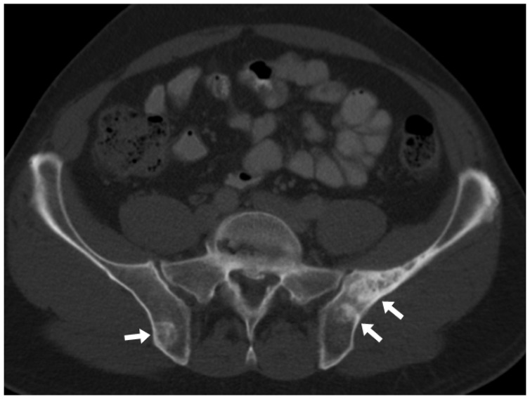
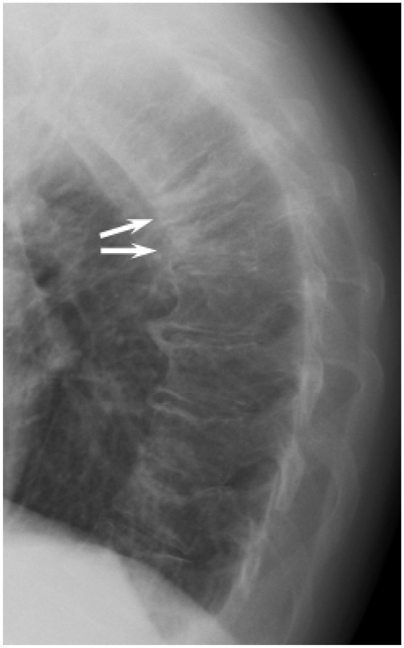
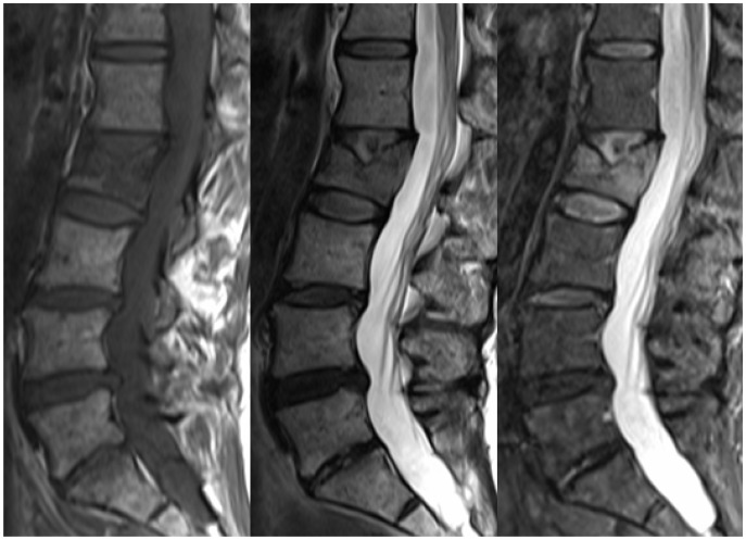
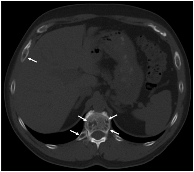
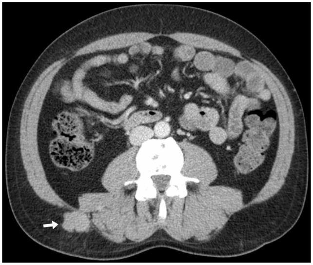
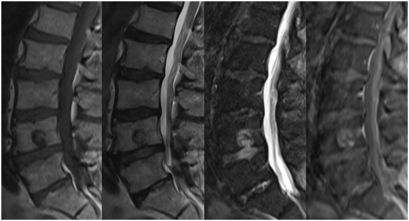
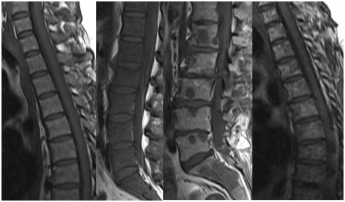
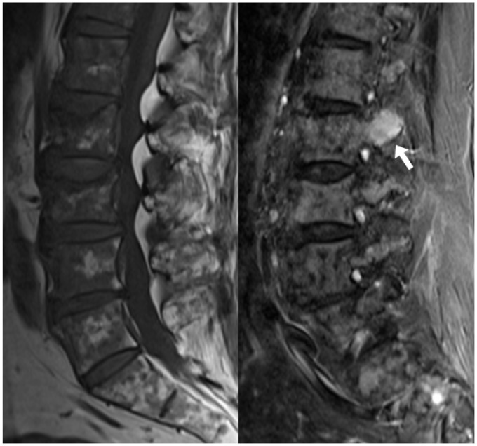
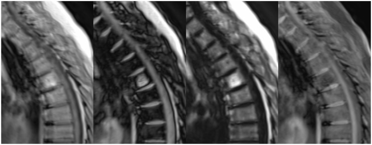
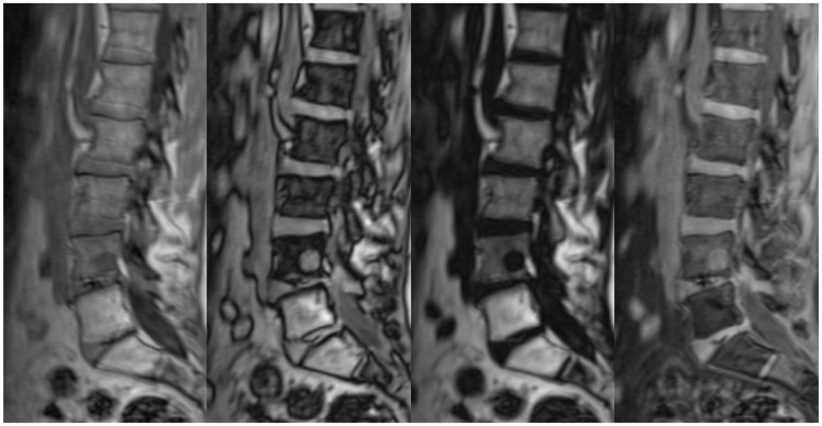
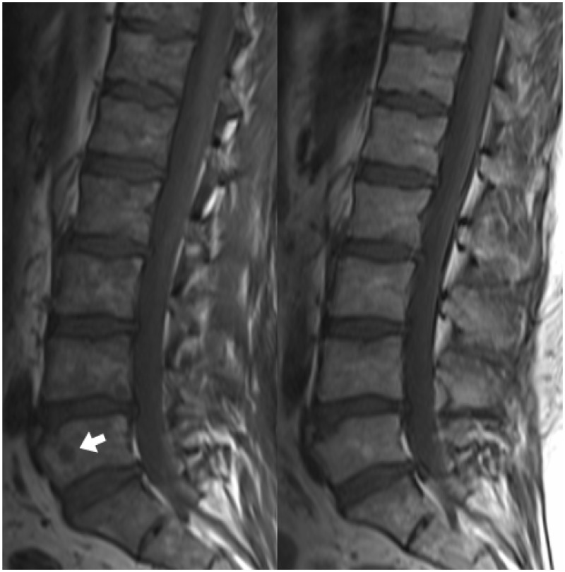
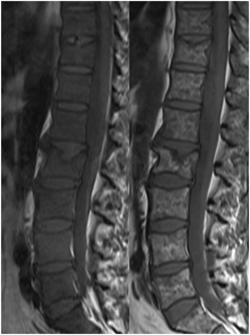
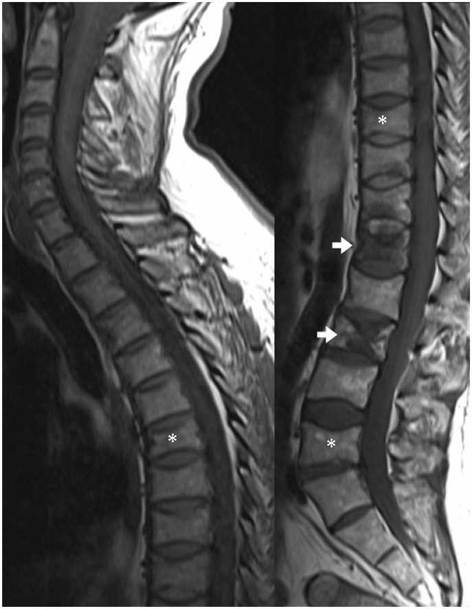
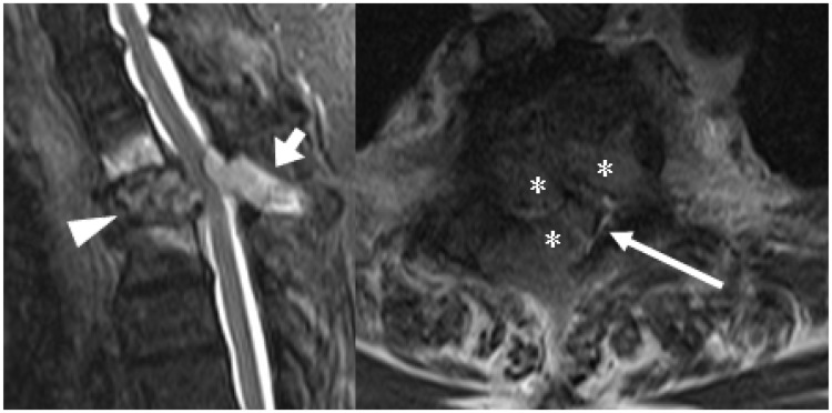
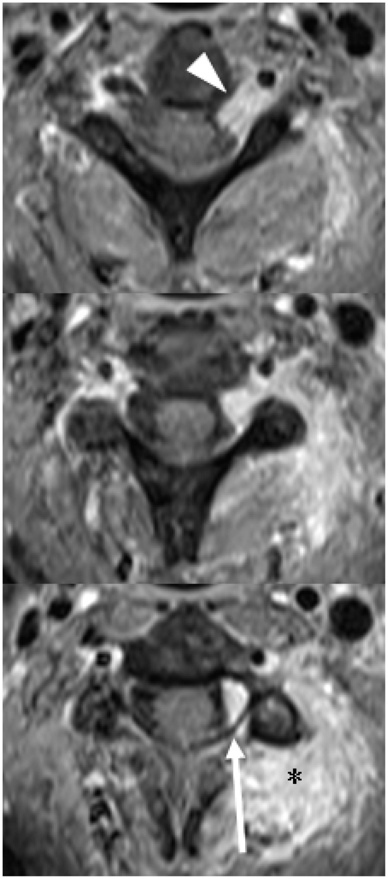
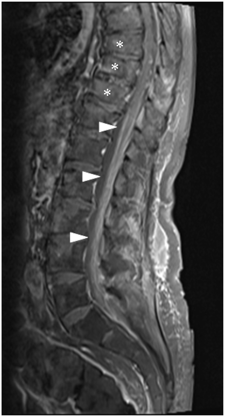
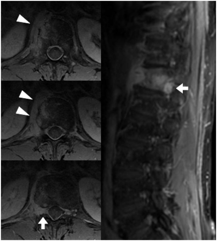
Radiology provides a crucial clinical adjunct in patients with plasma cell disorders, in particular multiple myeloma, and its uses are evolving and expanding. This pictorial review illustrates the role of imaging throughout the patient's clinical course, with specific reference to recently updated international diagnostic criteria. At presentation, imaging optimises characterisation and staging of the plasma-cell disorder, while later in the course of the disease, its roles include the monitoring of disease progression, assessment of post-treatment response and the investigation of clinical deterioration.

Keywords: Magnetic resonance imaging; multiple myeloma; plasmacytoma.


Figures

Figure 1.
Figure 1.
Axial CT scan on bone windows showing multiple sclerotic lesions in the bony pelvis in a patient with osteosclerotic myeloma. CT: computed tomography.
Figure 2.
Figure 2.
Lateral chest X-ray showing two contiguous thoracic vertebral crush fractures (arrows). The visualised spine is osteopaenic, but there are no definite discrete lesions.
Figure 3.
Figure 3.
Sagittal T1, T2 and T2 STIR images of the lumbar spine showing a pathological crush fracture of the L2 vertebral body. The diffuse T1 hypointensity of the vertebral body indicates an underlying lesion, consistent with a plasmacytoma given known MM. MM: multiple myeloma.
Figure 4.
Figure 4.
Axial CT of the upper abdomen on bone windows demonstrating multiple lytic vertebral and rib lesions (arrows). CT: computed tomography.
Figure 5.
Figure 5.
Axial CT on soft-tissue windows shows a subcutaneous mass in the right flank adjacent to the paravertebral musculature (arrowhead), proven to represent an extramedullary plasmacytoma on biopsy. CT: computed tomography.
Figure 6.
Figure 6.
Sagittal T1, T2, T2 STIR and T1 post-contrast with fat saturation images demonstrating a plasmacytoma in the L4 vertebral body. The mass is hypointense on the T1 and T2 images, hyperintense on the T2 STIR sequence and demonstrates enhancement. The background marrow appearances are normal.
Figure 7.
Figure 7.
Sagittal T1 images showing possible appearances of the vertebral marrow: normal, diffuse infiltration, discrete lesions and a salt-and-pepper appearance.
Figure 8.
Figure 8.
The sagittal T1 image of the lumbar spine shows a salt-and-pepper appearance of the vertebral marrow in a patient with MM, obscuring discrete lesions. The T2 STIR sequence, however, reveals a discrete hyperintense mass within the left posterior elements of L2 (arrow), consistent with a plasmacytoma. MM: multiple myeloma.
Figure 9.
Figure 9.
Sagittal in-phase, out-of-phase, fat-selective and water-selective images of a haemangioma within the T6 vertebral body. Most notably, the lesion shows high signal on the fat-selective image but low signal on the water-selective image.
Figure 10.
Figure 10.
Sagittal in-phase, out-of-phase, fat-selective and water-selective images of a plasmacytoma within the L4 vertebral body. In contrast to Figure 9, the lesion shows low signal on the fat-selective image and high signal on the water-selective image. Note the fatty marrow signal (high signal on the fat-selective image) in the L5 vertebral body and sacrum due to radiotherapy for management of a previous sacral plasmacytoma.
Figure 11.
Figure 11.
Sagittal T1 images of the lumbar spine in a patient with MM, pre and post treatment. Before treatment, the T1 signal is heterogeneous and generally decreased. There is also a small T1-hypointense lesion in the L5 vertebral body (arrow). After treatment, the T1 signal has normalised, and the L5 lesion is no longer visible. MM: multiple myeloma.
Figure 12.
Figure 12.
Sagittal T1 images pre and post treatment in another patient. Before treatment, the T1 signal is diffusely decreased, consistent with diffuse bone-marrow infiltration. After treatment, the appearances have altered to a ‘salt-and-pepper’ pattern. Crush fractures of the T11 and L2 vertebral bodies are also present.
Figure 13.
Figure 13.
Sagittal T1 images of the upper and lower spine in a patient with multiple vertebral plasmacytomas. Multiple vertebral crush fractures are present, some due to underlying bone lesions (arrows) and others without discrete lesions (asterisks).
Figure 14.
Figure 14.
The sagittal STIR image (left) in a patient with leg symptoms shows a plasmacytoma involving both the anterior (arrowhead) and posterior elements (short arrow) of the T2 vertebra. The axial T2 image (right) confirms the presence of tumour extending into the central vertebral canal (asterisks), compressing and distorting the spinal cord (long arrow), but without cord signal change.
Figure 15.
Figure 15.
Contiguous axial post-contrast T1 images (superior to inferior) showing extensive extra-osseous tumour (asterisk), including extension into the left C2/3 neural exit foramen (arrowhead). There is also some tumour within the central vertebral canal (long arrow), but without distortion of the spinal cord or cord compression.
Figure 16.
Figure 16.
Sagittal post-contrast T1 with fat saturation image of the lumbar spine demonstrating leptomeningeal infiltration (arrowheads) involving the lower spinal cord, conus medullaris and cauda equina. Pathological vertebral body crush fractures are also present (asterisks).
Figure 17.
Figure 17.
Axial and coronal post-contrast T1 with fat saturation images show a vertebral plasmacytoma with extra-osseous extension (arrowheads), suspected to be producing dural involvement within the neural exit foramen (short arrows). This patient had proven meningeal myelomatosis on lumbar puncture.

Similar articles

Cited by

References

    1. Rajkumar SV, Dimopoulos MA, Palumbo A, et al. International Myeloma Working Group updated criteria for the diagnosis of multiple myeloma. Lancet Oncol 2014; 15: e538–e548. - PubMed
    1. Bird JM, Owen RG, D’Sa S, et al. Guidelines for the diagnosis and management of multiple myeloma 2011. Br J Haematol 2011; 154: 32–75. - PubMed
    1. Durie BGM, Kyle RA, Belch A, et al. Myeloma management guidelines: a consensus report from the Scientific Advisors of the International Myeloma Foundation. Hematol J 2003; 4: 379–398. - PubMed
    1. Soutar R, Lucraft H, Jackson G, et al. Guidelines on the diagnosis and management of solitary plasmacytoma of bone and solitary extramedullary plasmacytoma. Br J Haematol 2004; 124: 717–726. - PubMed
    1. Moulopoulos LA, Dimopoulos MA, Weber D, et al. Magnetic resonance imaging in the staging of solitary plasmacytoma of bone. J Clin Oncol 1993; 11: 1311–1315. - PubMed

MeSH terms

LinkOut - more resources