

Anterior spinal artery aneurysm in aortic stenosis of different etiology: Report of three cases
- PMID: 28424013
- PMCID: PMC5433591
- DOI: 10.1177/1971400917690008
Anterior spinal artery aneurysm in aortic stenosis of different etiology: Report of three cases
Abstract
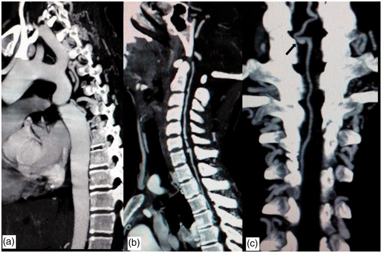
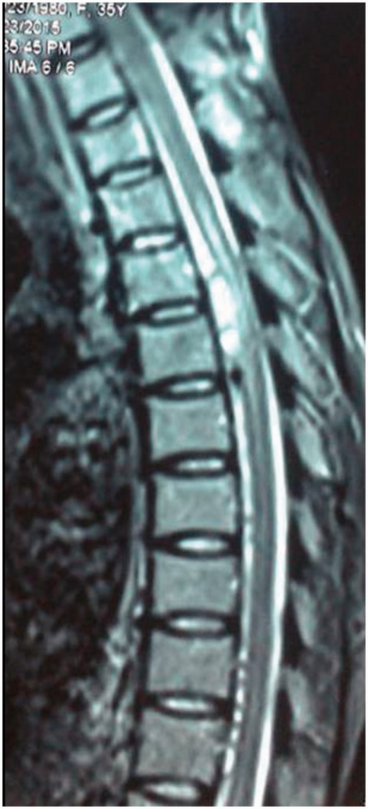
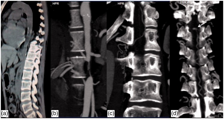
Isolated aneurysms of spinal arteries are rare. Spinal artery aneurysms are commonly found in association with spinal cord arteriovenous malformation and coarctation of aorta and rarely with aortic arch interruption and Klippel-Trenaunay syndrome. Spinal angiograms are the gold standard for diagnosing these spinal artery aneurysms but with the advances in computed tomography technology these aneurysms can also be very well demonstrated in computed tomography angiograms. We describe three cases of anterior spinal artery aneurysm, those are flow related aneurysms, associated with coarctation of aorta and with Takayasu arteritis.
Keywords: Anterior spinal artery; Takayasu arteritis; aneurysm; coarctation of aorta; computed tomography angiography.
Figures


Similar articles
-
Ruptured anterior spinal artery aneurysm: a case report.Surg Neurol. 1999 Jun;51(6):608-12. doi: 10.1016/s0090-3019(98)00114-1. Surg Neurol. 1999. PMID: 10369227 Review.
-
Successful Surgical Resection of Spinal Artery Aneurysms: Report of 3 Cases.World Neurosurg. 2018 Jan;109:171-178. doi: 10.1016/j.wneu.2017.09.174. Epub 2017 Oct 4. World Neurosurg. 2018. PMID: 28987836
-
Cervical spine dural arteriovenous fistula with coexisting spinal radiculopial artery aneurysm presenting as subarachnoid hemorrhage: case report.Neurosurgery. 2012 Jan;70(1):E259-63; discussion E263. doi: 10.1227/NEU.0b013e31822ac0fb. Neurosurgery. 2012. PMID: 21795862
-
Ruptured anterior spinal artery aneurysm associated with coarctation of aorta. Case report and literature review.Interv Neuroradiol. 2002 Sep 30;8(3):285-92. doi: 10.1177/159101990200800308. Epub 2004 Oct 20. Interv Neuroradiol. 2002. PMID: 20594486 Free PMC article.
-
Ruptured spinal artery aneurysm associated with coarctation of the aorta.World Neurosurg. 2014 Feb;81(2):441.e17-22. doi: 10.1016/j.wneu.2012.07.027. Epub 2012 Aug 7. World Neurosurg. 2014. PMID: 22885167 Review.
Cited by
-
Isolated spinal aneurysms with spontaneous regression.Neurosurg Rev. 2025 Sep 8;48(1):635. doi: 10.1007/s10143-025-03768-8. Neurosurg Rev. 2025. PMID: 40920243 Free PMC article.
-
Ruptured isolated spinal artery aneurysms: a rare manifestation of an arterial dissecting disease.Front Neurol. 2025 May 29;16:1567536. doi: 10.3389/fneur.2025.1567536. eCollection 2025. Front Neurol. 2025. PMID: 40510201 Free PMC article. Review.
-
Diagnostic and Therapeutic Approaches for Spinal Subarachnoid Hemorrhage Due to Spinal Aneurysms and Other Etiologies.J Clin Med. 2025 Mar 31;14(7):2398. doi: 10.3390/jcm14072398. J Clin Med. 2025. PMID: 40217848 Free PMC article.
-
Management and outcomes for thoracic anterior spinal artery aneurysms: illustrative case.J Neurosurg Case Lessons. 2025 Jun 23;9(25):CASE24649. doi: 10.3171/CASE24649. Print 2025 Jun 23. J Neurosurg Case Lessons. 2025. PMID: 40550205 Free PMC article.
-
Ruptured isolated spinal artery aneurysms: Case series of five patients and a review of the literature on management strategies.Interv Neuroradiol. 2025 Jun;31(3):402-413. doi: 10.1177/15910199221149562. Epub 2023 Jan 10. Interv Neuroradiol. 2025. PMID: 36628492 Free PMC article. Review.
References
-
- Rengachary SS, Duke DA, Tsai FY, et al. Spinal arterial aneurysm: Case report. Neurosurgery 1993; 33: 125–129. - PubMed
-
- Smith BS, Penka CF, Erickson LS, et al. Subarachnoid hemorrhage due to anterior spinal artery aneurysm. Neurosurgery 1986; 18: 217–219. - PubMed
-
- Singh V, Naik S, Shetty GS, et al. Anterior spinal artery aneurysm in a case of Moyamoya disease. Acta Neurol Belg 2016; 116: 663–665. - PubMed
-
- Chen CC, Bellon RJ, Ogilvy CS, et al. Aneurysms of the lateral spinal artery: Report of two cases. Neurosurgery 2001; 48: 949–953. - PubMed
Publication types
MeSH terms
LinkOut - more resources
Full Text Sources
Other Literature Sources
Medical
