. 2017 May 15;198(10):3939-3948.
doi: 10.4049/jimmunol.1601078. Epub 2017 Apr 19.

mTORC1 Promotes T-bet Phosphorylation To Regulate Th1 Differentiation

Affiliations

mTORC1 Promotes T-bet Phosphorylation To Regulate Th1 Differentiation

Olesya Chornoguz et al. J Immunol. .

Abstract

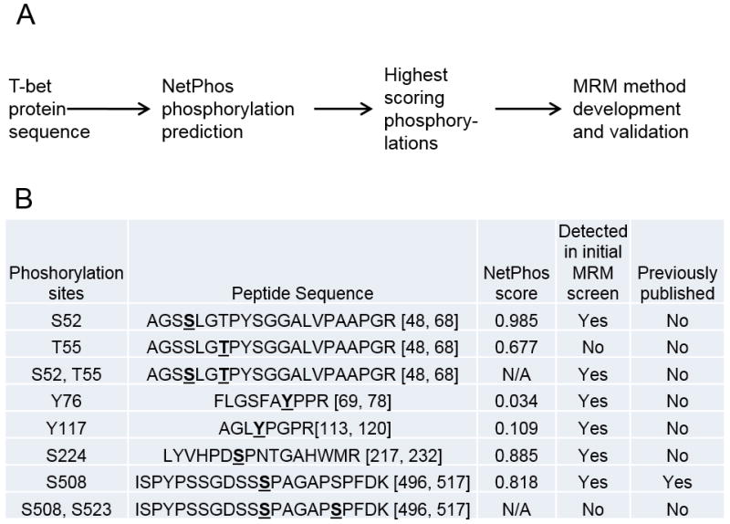
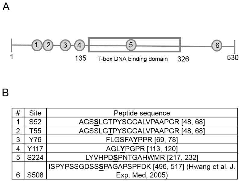
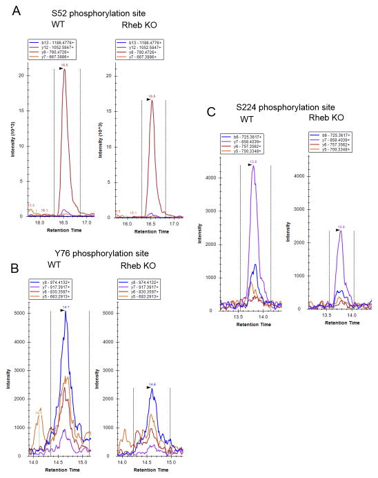
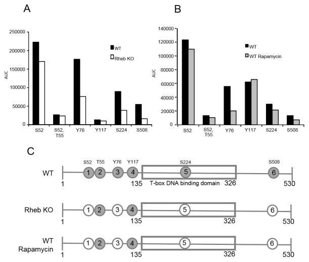
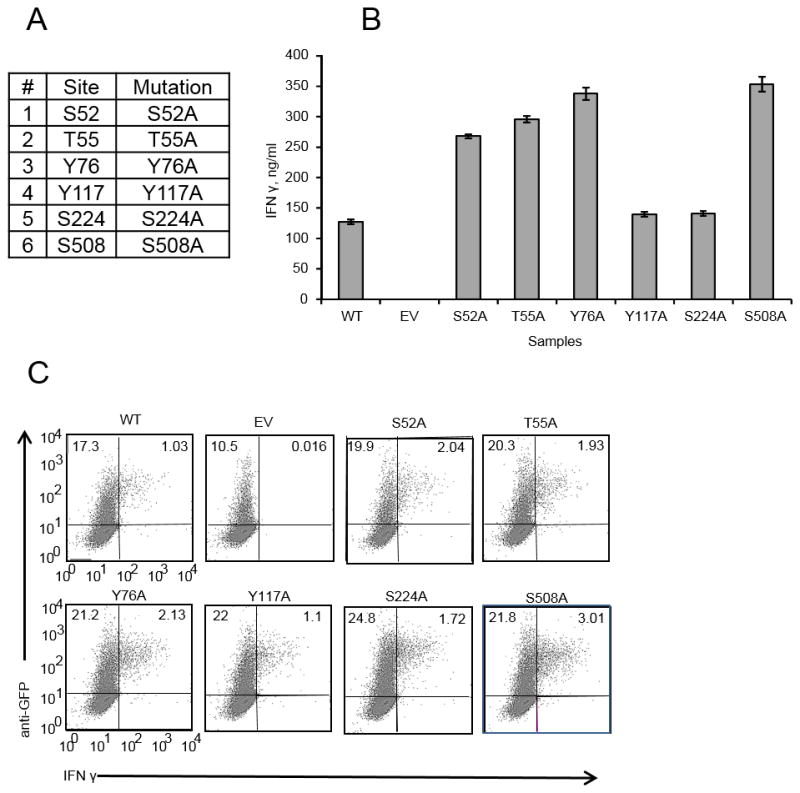
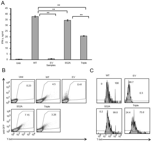
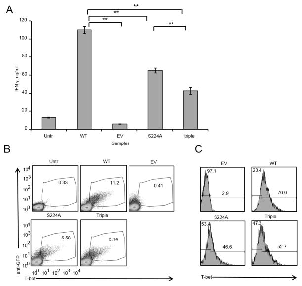
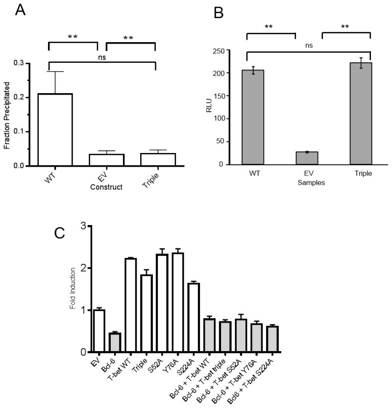
CD4+ T cells lacking the mTORC1 activator Rheb fail to secrete IFN-γ under Th1 polarizing conditions. We hypothesized that this phenotype is due to defects in regulation of the canonical Th1 transcription factor T-bet at the level of protein phosphorylation downstream of mTORC1. To test this hypothesis, we employed targeted mass-spectrometry proteomic analysis-multiple reaction monitoring mass spectrometry. We used this method to detect and quantify predicted phosphopeptides derived from T-bet. By analyzing activated murine wild-type and Rheb-deficient CD4+ T cells, as well as murine CD4+ T cells activated in the presence of rapamycin, a pharmacologic inhibitor of mTORC1, we were able to identify six T-bet phosphorylation sites. Five of these are novel, and four sites are consistently dephosphorylated in both Rheb-deficient CD4+ T cells and T cells treated with rapamycin, suggesting mTORC1 signaling controls their phosphorylation. Alanine mutagenesis of each of the six phosphorylation sites was tested for the ability to impair IFN-γ expression. Single phosphorylation site mutants still support induction of IFN-γ expression; however, simultaneous mutation of three of the mTORC1-dependent sites results in significantly reduced IFN-γ expression. The reduced activity of the triple mutant T-bet is associated with its failure to recruit chromatin remodeling complexes to the Ifng gene promoter. These results establish a novel mechanism by which mTORC1 regulates Th1 differentiation, through control of T-bet phosphorylation.


Figures

Figure 1
Figure 1. Proteomic workflow for discovery and validation of novel T-bet phosphorylation sites
A, Mouse T-bet protein sequence was used as an input for phosphorylation site prediction analysis using NetPhos program. T-bet phosphorylations with the highest predicted scores were chosen for LC-MRM-MS method development. B, Phosphorylation sites and their NetPhos scores that were chosen for preliminary MRM method development.
Figure 2
Figure 2. Six phosphorylation sites were detected and validated using MRM-MS
A, Schematic of T-bet protein with T-box DNA binding domain shown as a gray rectangle in the middle. The numbers in gray circles show the location of the six different phosphorylations on the T-bet sequence. B, List of the six phosphorylations detected using MRM-MS and their corresponding tryptic peptides.
Figure 3
Figure 3. LC-MRM-MS traces of T-bet peptides that contain phosphorylations regulated by mTORC1
Different color traces represent different fragments of the same peptide. The fragments are denoted at the top of each LC-MRM-MS trace. A, S52 phosphorylation site, peptide AGSSLGTPYSGGALVPAAPGR, B, Y76 phosphorylation site, peptide FLGSFAYPPR, C, S224 phosphorylations site, peptide LYVHPDSPNTGAHWMR. Data are representative of 5 individual experiments.
Figure 4
Figure 4. Three of the novel T-bet phosphorylations are mTORC1 dependent
A, Wild-type C57/BL6 (WT) or Rhebfl/fl CD4+Cre (Rheb KO) CD4+ T cells were purified from spleens and lymph nodes of 6–12 week old mice and activated with plate-bound anti-CD3 and soluble anti-CD28 for 24 hours in Th1 polarizing culture conditions. Activated T cells were harvested, lysed with Rapigest, trypsin digested and analyzed by LC-MRM-MS. AUC on the y axis is area under the curve – measure of phosphorylated peptide abundance. Data are from one of 2 experiments with T cells from individual mice. B, CD4+ T cells were purified from spleens and lymph nodes of WT mice and activated for 48 hours as in A in the presence or absence of 500 nM Rapamycin (WT or WT Rapamycin), prepared and analyzed as in A. Data are from one of 3 experiments with T cells from individual mice. C, Schematic of T-bet with phosphorylations shown as circles with numbers. Gray circles are the phosphorylations sites. White circles show phosphorylations that are mTORC1 dependent. Data are from one of 5 experiments with T cells from individual mice.
Figure 5
Figure 5. Single phosphorylation site T-bet mutants are not deficient in their ability to induce IFNγ
A, Six single phosphorylation site mutants. All of the phosphorylation sites were mutated to alanines. B, EL4 cells were transfected with either WT, empty vector (EV) or different mutants. 24 hours post-transfection equal numbers of GFP+ cells were plated and activated with PMA/Ionomycin overnight. Supernatants were analyzed for IFNγ by ELISA. Error bars are Standard Error of the Mean (SEM). C, EL4 cells transfected as in B and stimulated with PMA/Ionomycin overnight in the presence of GolgiPlug protein transport inhibitor. Data are from one of 3 experiments.
Figure 6
Figure 6. Triple phosphorylation site T-bet mutant has a significantly reduced ability to induce IFNγ in EL4
A. EL4 cells were transfected with either WT, empty vector, S52A single phosphorylation site mutant or triple phosphorylation site mutant. 24 hours post-transfection equal numbers of GFP+ cells were plated and activated with PMA/Ionomycin overnight. Supernatants were analyzed for IFNγ by ELISA. Error bars are SEM, ** P<0.05 by 1-way ANOVA with Bonferroni’s Multiple Comparison Test. B, EL4 transfected as in A were fixed, permeabilized and stained with anti-GFP and anti-T-bet antibodies. C, Samples from B gated on GFP+ cells and T-bet levels are shown as histograms. Data are representative of 4 experiments.
Figure 7
Figure 7. Triple phosphorylation site T-bet mutant has a significantly reduced ability to induce IFNγ in T-bet deficient primary CD4+ T cells
A. CD4+ T cells were purified from spleens and lymph nodes of T-bet deficient (T-bet KO) mice and transduced with either WT, empty vector, S224A single phosphorylation site mutant or triple mutant construct containing retroviruses. 48 hours post-transduction equal numbers of GFP+ cells were plated and activated with 1 μg/ml of plate-bound anti-CD3, 2 μg/ml of soluble anti-CD28 overnight. Supernatants were analyzed for IFNγ by ELISA. Error bars are SEM; ** P<0.05 by 1-way ANOVA with Bonferroni’s Multiple Comparison Test. B, T-bet KO CD4+ T cells transduced with the constructs indicated in A were fixed, permeabilized and stained with anti-GFP and anti-T-bet antibodies. C, Samples from B gated on GFP+ cells and T-bet levels are shown as histograms. Data are from one of 2 experiments with T cells from 4 individual mice.
Figure 8
Figure 8. Triple mutant T-bet has a reduced ability to promote addition of permissive chromatin remodeling mark H3K4me2 but can still bind to the Ifng and SOCS3 promoters
A. EL4 cells were transiently transfected with empty vector (EV), WT T-bet, or T-bet triple mutant, and chromatin was fixed after 72 hours and sonicated for ChIP. Graph depicts fraction of input DNA precipitated by anti H3K4me2 specific antibody from each sample, mean ± SD, N=3 replicates. ** P<0.05 by 1-way ANOVA with Bonferroni’s Multiple Comparison Test. Control rabbit IgG precipitated a negligible amount of input and is not shown. The experiment was performed 3 times with similar results. B. EL4 cells were co-transfected with the pGL3 Ifng promoter reporter construct, pRLtk Renilla control, and either WT T-bet, triple mutant T-bet or empty vector (EV). After PMA/Ionomycin stimulation, firefly luciferase activity was normalized to the activity of co-transfected Renilla control. RLU – relative luminescence units. Error bars are SD; **P<0.05 by 1-way ANOVA with Bonferroni’s Multiple Comparison Test. Data are from one of 2 experiments. C. EL4 were co-transfected with the SOCS3 luciferase promoter reporter construct and empty vector (EV) or T-bet constructs in the absence (open bars) or the presence of Bcl-6 (grey bars). After PMA/Ionomycin stimulation, firefly luciferase activity was normalized to co-transfected Renilla luciferase activity and is depicted as fold induction compared to EV. Error bars depict SD, and data are representative of 4 independent experiments.

References

    1. Wan YY. Multi-tasking of helper T cells. Immunology. 2010;130:166–171. - PMC - PubMed
    1. Lucas S, Ghilardi N, Li J, de Sauvage FJ. IL-27 regulates IL-12 responsiveness of naive CD4++ T cells through Stat1-dependent and -independent mechanisms. Proceedings of the National Academy of Sciences of the United States of America. 2003;100:15047–15052. - PMC - PubMed
    1. Szabo SJ, Kim ST, Costa GL, Zhang X, Fathman CG, Glimcher LH. A novel transcription factor, T-bet, directs Th1 lineage commitment. Cell. 2000;100:655–669. - PubMed
    1. Lazarevic V, Glimcher LH. T-bet in disease. Nature immunology. 2011;12:597–606. - PMC - PubMed
    1. Delgoffe GM, Kole TP, Zheng Y, Zarek PE, Matthews KL, Xiao B, Worley PF, Kozma SC, Powell JD. The mTOR kinase differentially regulates effector and regulatory T cell lineage commitment. Immunity. 2009;30:832–844. - PMC - PubMed

MeSH terms