

Phosphorylation Induces Structural Changes in the Autographa californica Nucleopolyhedrovirus P10 Protein
- PMID: 28424279
- PMCID: PMC5469262
- DOI: 10.1128/JVI.00002-17
Phosphorylation Induces Structural Changes in the Autographa californica Nucleopolyhedrovirus P10 Protein
Abstract
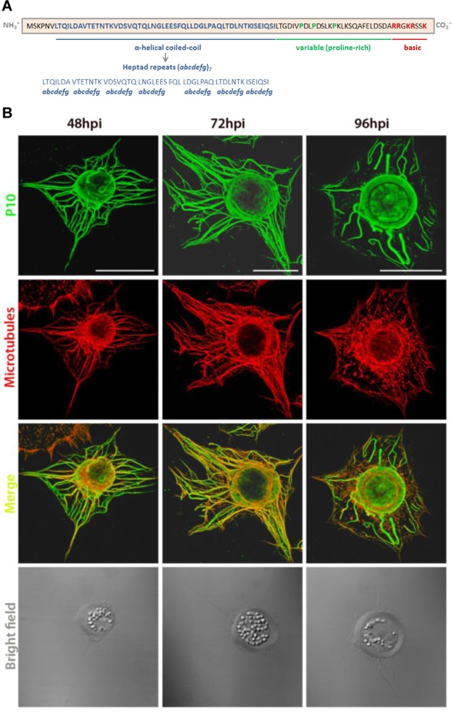
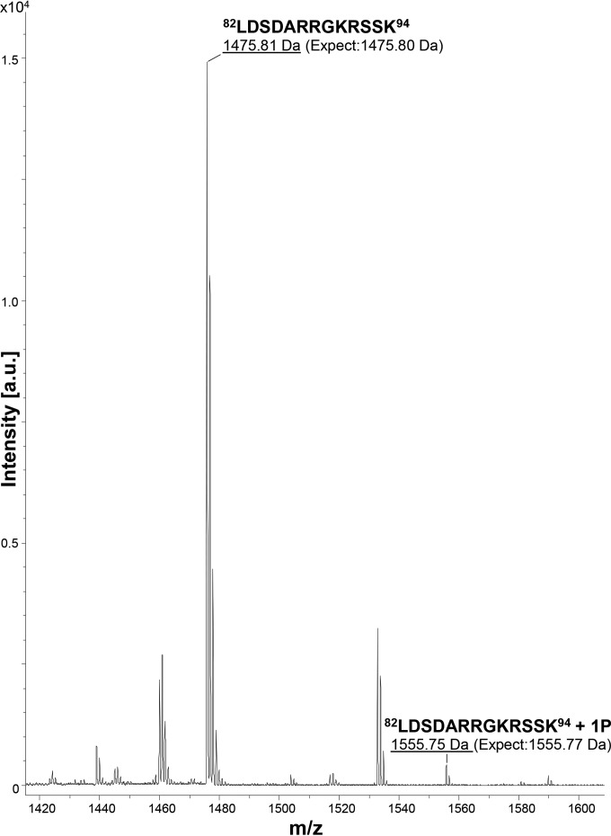
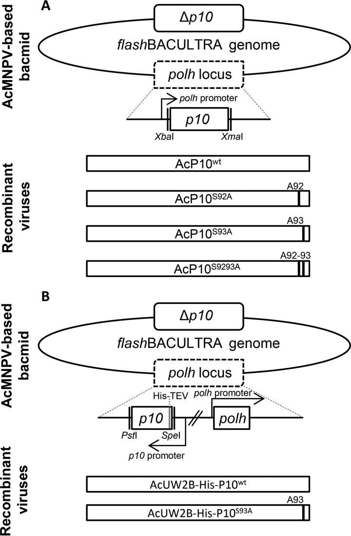
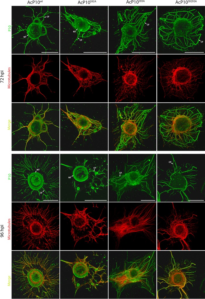
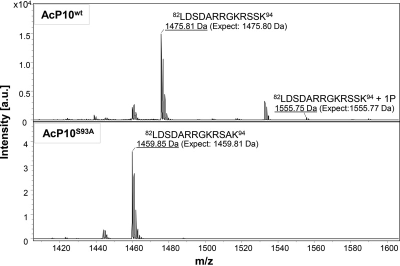
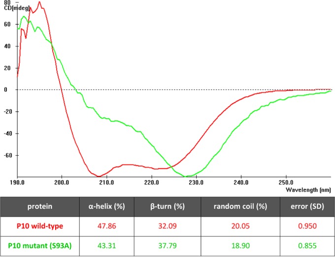
Baculoviruses encode a variety of auxiliary proteins that are not essential for viral replication but provide them with a selective advantage in nature. P10 is a 10-kDa auxiliary protein produced in the very late phase of gene transcription by Autographa californica multiple nucleopolyhedrovirus (AcMNPV). The P10 protein forms cytoskeleton-like structures in the host cell that associate with microtubules varying from filamentous forms in the cytoplasm to aggregated perinuclear tubules that form a cage-like structure around the nucleus. These P10 structures may have a role in the release of occlusion bodies (OBs) and thus mediate the horizontal transmission of the virus between insect hosts. Here, using mass spectrometric analysis, it is demonstrated that the C terminus of P10 is phosphorylated during virus infection of cells in culture. Analysis of P10 mutants encoded by recombinant baculoviruses in which putative phosphorylation residues were mutated to alanine showed that serine 93 is a site of phosphorylation. Confocal microscopy examination of the serine 93 mutant structures revealed aberrant formation of the perinuclear tubules. Thus, the phosphorylation of serine 93 may induce the aggregation of filaments to form tubules. Together, these data suggest that the phosphorylation of serine 93 affects the structural conformation of P10.IMPORTANCE The baculovirus P10 protein has been researched intensively since it was first observed in 1969, but its role during viral infection remains unclear. It is conserved in the alphabaculoviruses and expressed at high levels during virus infection. Producing large amounts of a protein is wasteful for the virus unless it is advantageous for the survival of its progeny, and therefore, P10 presents an enigma. As P10 polymerizes to form organized cytoskeletal structures that colocalize with host cell microtubules, the structural relationship of the protein with the host cell may present a key to help understand the function and importance of this protein. This study addresses the importance of the structural changes in P10 during infection and how they may be governed by phosphorylation. The P10 structures affected by phosphorylation are closely associated with the viral progeny and thus may potentially be responsible for its dissemination and survival.
Keywords: AcMNPV; P10; Trichoplusia ni; baculovirus; cytoskeleton; host-cell interactions; microtubules; occlusion bodies; protein phosphorylation.
Copyright © 2017 American Society for Microbiology.
Figures
Similar articles
-
The baculovirus P10 protein of Autographa californica nucleopolyhedrovirus forms two distinct cytoskeletal-like structures and associates with polyhedral occlusion bodies during infection.Virology. 2008 Feb 20;371(2):278-91. doi: 10.1016/j.virol.2007.09.043. Epub 2007 Nov 8. Virology. 2008. PMID: 17991504
-
The heptad repeats region is essential for AcMNPV P10 filament formation and not the proline-rich or the C-terminus basic regions.Virology. 2007 Sep 1;365(2):390-7. doi: 10.1016/j.virol.2007.03.051. Epub 2007 May 3. Virology. 2007. PMID: 17481692
-
Formation of P10 tubular structures during AcMNPV infection depends on the integrity of host-cell microtubules.Virology. 2003 Dec 20;317(2):308-20. doi: 10.1016/j.virol.2003.08.035. Virology. 2003. PMID: 14698669
-
Post-translational modification of baculovirus-encoded proteins.Virus Res. 2020 Apr 2;279:197865. doi: 10.1016/j.virusres.2020.197865. Epub 2020 Jan 24. Virus Res. 2020. PMID: 31987850 Review.
-
[Structure and Function of the Baculovirus Per Os Infectivity Factor (PIF) P74].Bing Du Xue Bao. 2016 Jul;32(4):523-8. Bing Du Xue Bao. 2016. PMID: 29996043 Review. Chinese.
Cited by
-
Label-free proteomics analysis on the envelope of budded viruses of Bombyx mori nucleopolyhedrovirus harboring differential localized GP64.Virus Genes. 2023 Apr;59(2):260-275. doi: 10.1007/s11262-022-01961-1. Epub 2022 Dec 13. Virus Genes. 2023. PMID: 36512182
-
In cultured cells the baculovirus P10 protein forms two independent intracellular structures that play separate roles in occlusion body maturation and their release by nuclear disintegration.PLoS Pathog. 2019 Jun 10;15(6):e1007827. doi: 10.1371/journal.ppat.1007827. eCollection 2019 Jun. PLoS Pathog. 2019. PMID: 31181119 Free PMC article.
-
Dissecting the Cell Entry Pathway of Baculovirus by Single-Particle Tracking and Quantitative Electron Microscopic Analysis.J Virol. 2019 Apr 3;93(8):e00033-19. doi: 10.1128/JVI.00033-19. Print 2019 Apr 15. J Virol. 2019. PMID: 30760565 Free PMC article.
References
-
- Federici BA. 1986. Ultrastructure of baculoviruses, p 61–88. In Granados, RR, Federici, BA (ed), The biology of baculoviruses. CRC Press, Boca Raton, FL.
-
- Bilimoria S. 1986. Taxonomy and identification of baculoviruses, p 37–59. In Granados R, Federeci B (ed), The biology of baculoviruses. CRC Press, Boca Raton, FL.
Publication types
MeSH terms
Substances
LinkOut - more resources
Full Text Sources
Other Literature Sources
Research Materials
Miscellaneous
