. 2017 Apr 19;7(1):960.
doi: 10.1038/s41598-017-00922-1.

A lack of coordination between sister-chromatids segregation and cytokinesis in the oocytes of B6.YTIR (XY) sex-reversed female mice

Affiliations

A lack of coordination between sister-chromatids segregation and cytokinesis in the oocytes of B6.YTIR (XY) sex-reversed female mice

Jia-Qiao Zhu et al. Sci Rep. .

Abstract

The B6.YTIR (XY) mouse develops bilateral ovaries despite the expression of the testis-determining gene Sry during gonadal differentiation. We reported that the oocytes of the XY female are defective in their cytoplasm, resulting in a failure in the second meiotic division after activation or fertilization in vitro. However, the mechanism of meiotic failure or the cause of infertility remained to be clarified. In the present study, we obtained mature oocytes from XY females by superovulation and confirmed that these oocytes also fail in zygotic development. By using confocal microscopy 3D-analysis, we demonstrated that meiotic spindles were properly positioned and oriented in the MII-oocytes from XY females. After parthenogenic activation, fewer oocytes from XY females extruded the second polar body, and in those oocytes, sister-chromatids were often separated but neither set entered the second polar body. ARP2, F-actin, and ORC4, known to play roles in asymmetric meiotic division, were initially localized along the ooplasmic membrane and concentrated over the MII-spindle but lost their cortical polarity after activation while the sister-chromatids moved away from the oolemma in the oocytes from XY females. Our results indicate that the second polar body extrusion is uncoupled from the sister-chromatids separation in the oocytes from XY female mouse.


Conflict of interest statement

The authors declare that they have no competing interests.

Figures

Figure 1
Figure 1
eCG-dosage dependent ovulation of oocytes from XX and XY females. The number of ovulated oocytes was larger after injection with 10 IU eCG than 5 IU eCG in both XX and XY females. The number of ovulated oocytes was also larger from XX females than XY females with 10 IU eCG. Data are presented as means ± SEM. The total number of females examined is given in parentheses at the top of each column. * and ** significant differences at p < 0.05 and 0.001, respectively.
Figure 2
Figure 2
Failure in the embryonic development from the oocytes ovulated by XY females. Each column indicates the percentages of embryos at different stages at 1–5 days after IVF. “Dead” includes dead or fragmented embryos. The total number of oocytes examined is given in parentheses at the top of each graph.
Figure 3
Figure 3
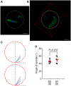
Normal orientation of MII-spindles in the IVM-oocytes from XY females. (A) 2D-image of a single z-section. An MII-oocyte from an XY female stained with anti-α-tubulin antibody (green) and DAPI (blue). The white broken circle indicates the position of zona pellucida. Bar: 20 µm. (B) 3D-image reconstructed from more than 40 z-sections at every 2.0 μm. The red box indicates the boundaries of 3D-image. The white broken circle indicates the position of zona pellucida. Bar: 20 µm. (The video of the 3D-image is given in Supplementary Fig. S1) (C) Summary of spindle orientation in the oocytes from XX and XY females. The red broken circle in each picture indicates the position of zona pellucida. The vertical axis was drawn to go through the center of each oocyte and the farther pole of the spindle. The spindle orientations in individual oocytes are shown in different colors. The angle of the spindle axis was measured from the horizontal axis in each oocyte. (D) Summery of spindle angles measured in the oocytes from XX and XY females. No difference is found between the two genotypes. Data are presented as means ± SEM. The total number of oocytes examined was given in parentheses at the bottom.
Figure 4
Figure 4
Higher frequency of aneuploidy in the MII-oocytes from XY females. (A) Examples of aneuploidy analyses. While almost all oocytes from XX females showed normal number (=20) of chromosomes, the oocytes from XY females showed variable numbers such as hypoploid (e.g. 19) or hyperploid (e.g., 21). The oocyte with the normal number of chromosomes often showed precocious separation of sister chromatids (PSSC). Bar: 10 µm. The areas around the chromatids indicated by (a) and (b) are magnified in the right panels. (B) Percentages of oocytes with normal (=20) and abnormal chromosome numbers. (C) Percentages of oocytes with PSSC. Data are shown as means ± SEM. * and ** significant differences at p < 0.05 and 0.01, respectively. The experiments were repeated at least three times each. For in vivo maturation, total 60 and 54 oocytes from XX and XY females, respectively, were examined. For IVM, total 95 and 73 oocytes from XX and XY females, respectively, were examined.
Figure 5
Figure 5
Meiotic progression and pronucleus formation in the MII-oocytes from XX and XY females following parthenogenic activation. (A) Time-lapse microscopy analysis of the second meiotic division. Chromosomes were stained with Hoechst 33342 (blue) at the end of time-lapse imaging. Five types of oocytes are shown; (1) a typical oocyte from an XX female with extrusion of the second polar body and single pronucleus formation. The oocytes from XY females are seen (2) without the second polar body extrusion and with two pronuclei formation, (3) with extrusion of the second polar body and two pronuclei formation, (4) without the second polar body extrusion and with multiple pronuclei formation, or (5) with symmetric cell division. Bar: 20 µm. (The time-lapse videos are given in Supplementary Fig. S2.1–S2.5) (B) Percentages of oocytes with the second polar body extrusion. (C) The time required for the oocyte to initiate the first polar body degradation, the second polar body extrusion, and pronuleus formation. (D) Percentages of oocytes that formed different numbers of pronuclei. Data are presented as mean ± SEM. * and **, significant differences between the oocytes from XX females and those from XY females at p < 0.05 and 0.001, respectively. The experiments were repeated at least three times each. For the in vivo maturation, total 67 and 63 oocytes from XX and XY females, respectively, were examined. For IVM, total 89 and 99 oocytes from XX and XY females, respectively, were examined.
Figure 6
Figure 6
Spindle movement and sister chromatids segregation in the MII-oocytes from XX and XY females following parthenogenic activation. Meiotic spindles and chromosomes were visualized by injection of β5-tubulin-GFP and H2B-mCherry mRNAs into the GV-stage oocytes, followed by IVM. The images of oocytes from XY females were compressed from Z-stacks in order to capture the entire chromatids or pronuclei. Bar: 20 µm. (The time-lapse videos were given in Supplementary Fig. S3.1–S3.3.) The experiments were repeated at least three times each. Total 7 oocytes (1 or 2 oocytes at a time) were tested for each genotype.
Figure 7
Figure 7
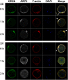
Loss of ORC4, ARP2 and F-actin polarization in the MII-oocytes from XY females after parthenogenic activation. ORC4, ARP2, and F-actin are localized along the oolemma and concentrated over the MII-spindle (arrows) in the ovulated-oocytes from both XX and XY females. ORC4 and F-actin are also concentrated but ARP2 is faint around the first polar body. After parthenogenic activation in the oocytes from XX females, all proteins are concentrated near the set of anaphase chromatids closer to oolemma (arrow) at 1 h and around the second polar body (arrow) at 2 h. In the oocyte from an XY female, by contrast, all proteins are localized evenly along the oolemma and, in addition, ARP2 has appeared in the ooplasm while the sister-chromatids are centered at 1 h after activation. In the oocyte at 2 h after activation, APR2 has disappeared from the oolemma entirely and distributed in the ooplasm while all ORC4, ARP2, and F-actin are concentrated around the set of anaphase chromatids near the oolemma. Bar: 20 µm. The experiments were repeated three times. Total 3, 10, and 10 oocytes of each genotype were examined at 0, 1, and 2 h, respectively.

Similar articles

Cited by

References

    1. Taketo T. The role of sex chromosomes in mammalian germ cell differentiation: can the germ cells carrying X and Y chromosomes differentiate into fertile oocytes? Asian J. Androl. 2015;17:360–6. - PMC - PubMed
    1. Yamamoto T-O. Progeny of artificially induced sex-reversals of male genotype (XY) in the medaka (Oryzias latipes) with special reference to YY-male. Genetics. 1955;40:406–19. - PMC - PubMed
    1. Espinosa MB, Vitullo AD. Offspring sex-ratio and reproductive performance in heterogametic females of the South American field mouse Akodon azarae. Reproduction in heterogametic Akodon azarae females. Hereditas. 1996;124:57–62. doi: 10.1111/j.1601-5223.1996.00057.x. - DOI - PubMed
    1. Hoekstra HE, Hoekstra JM. An unusual sex-determination system in south american field mice (genus AKODON): the role of mutation, selection, and meiotic drive in maintaining XY females. Evolution. 2001;55:190–197. doi: 10.1111/j.0014-3820.2001.tb01283.x. - DOI - PubMed
    1. Lovell-Badge R, Robertson E. XY female mice resulting from a heritable mutation in the primary testis-determining gene. Tdy. Development. 1990;109:635–646. - PubMed

Publication types

Grants and funding

LinkOut - more resources