. 2017 Apr 19;7(1):945.
doi: 10.1038/s41598-017-01006-w.

The glycogen synthase kinase MoGsk1, regulated by Mps1 MAP kinase, is required for fungal development and pathogenicity in Magnaporthe oryzae

Affiliations

The glycogen synthase kinase MoGsk1, regulated by Mps1 MAP kinase, is required for fungal development and pathogenicity in Magnaporthe oryzae

Tengsheng Zhou et al. Sci Rep. .

Erratum in

Abstract

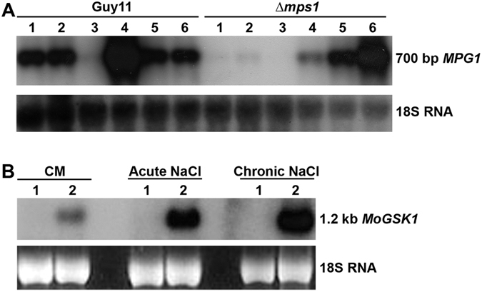
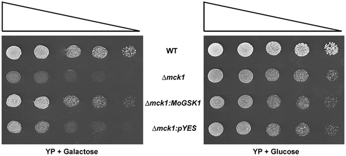
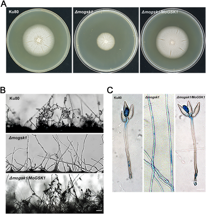
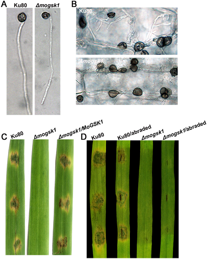
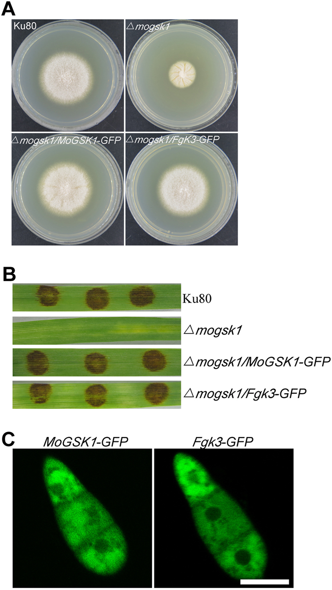
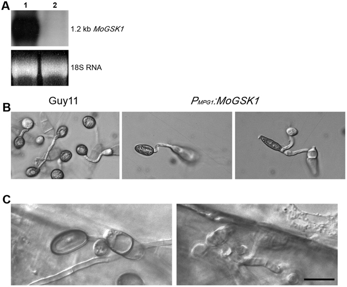
Magnaporthe oryzae, the causal agent of blast disease, is one of the most destructive plant pathogens, causing significant yield losses on staple crops such as rice and wheat. The fungus infects plants with a specialized cell called an appressorium, whose development is tightly regulated by MAPK signaling pathways following the activation of upstream sensors in response to environmental stimuli. Here, we show the expression of the Glycogen synthase kinase 3 (GSK3) MoGSK1 in M. oryzae is regulated by Mps1 MAP kinase, particularly under the stressed conditions. Thus, MoGSK1 is functionally characterized in this study. MoGsk1 is functionally homologues to the Saccharomyces cerevisiae GSK3 homolog MCK1. Gene replacement of MoGSK1 caused significant delay in mycelial growth, complete loss of conidiation and inability to penetrate the host surface by mycelia-formed appressorium-like structures, consequently resulting in loss of pathogenicity. However, the developmental and pathogenic defects of Δmogsk1 are recovered via the heterologous expression of Fusarium graminearum GSK3 homolog gene FGK3, whose coding products also shows the similar cytoplasmic localization as MoGsk1 does in M. oryzae. By contrast, overexpression of MoGSK1 produced deformed appressoria in M. oryzae. In summary, our results suggest that MoGsk1, as a highly conservative signal modulator, dictates growth, conidiation and pathogenicity of M. oryzae.


Conflict of interest statement

The authors declare that they have no competing interests.

Figures

Figure 1
Figure 1
Differential expression of MPG1 and GSK1 in the Δmps1 mutant compared to Guy11. (A) RNA gel blot analysis of MPG1 in Guy11 and Δmps1 in response to different growth conditions: CM medium (Lane 1), CM + 0.4 M NaCl (acute treatment) (Lane 2), CM + 0.4 M NaCl (chronic treatment) (Lane 3), MM medium − nitrate salts (Lane 4), MM − glucose (Lane 5), and sterile distilled water (Lane 6). (B) RNA gel blot analysis of MoGSK1 in Guy11 (Lane 1) and Δmps1 (Lane 2) in growth conditions of CM, CM + 0.4 M NaCl (acute treatment) and CM + 0.4 M NaCl (chronic treatment). Total RNA was extracted from mycelia cultured in CM for 48 hr then transferred to different liquid media indicated above for another 24 hr growth. For chronic NaCl treatment, mycelia were obtained after growth in CM + 0.4 M NaCl for 48 hr then transferred to fresh 0.4 M NaCl medium for another 24 hr growth.
Figure 2
Figure 2
M. oryzae MoGSK1 can functionally complement the Δmck1 mutant of S. cerevisiae. Yeast strains were grown at 12 °C for 14 days on galactose or glucose-supplemented YP medium. Plates were inoculated with 10 μl droplets containing 1 × 106, 5 × 105, 1 × 105, 5 × 104, or 1 × 104 cells/ml and left to grow for 14 days before examination. Triangles indicate the decreasing concentration of yeast cells. BY1471 (WT) is the wild type S. cerevisiae strain. Δmck1 is the yeast mutant sensitive to cold condition. The Δmck1:MoGSK1 transformant is expressing the M. oryzae MoGSK1 under the S. cerevisiae galactose-inducible GAL1 promoter. The negative control is the Δmck1 mutant transformed with the empty pYES vector.
Figure 3
Figure 3
Colony morphology and conidia formation of the Δmogsk1 mutant. (A) Colonies of Ku80, Δmogsk1 and Δmogsk3/MoGSK1 grown on complete medium (CM) at 26 °C. Photos were taken at 7 days after inoculation. (B) Comparison of conidia formation under light microscope between Ku80, Δmogsk1 and Δmogsk1/MoGSK1 after 48 hours induction at 26 °C on glass slides Bar = 50 μm. (C) Microscopic observation of aerial structures stained by lactophenol aniline blue. Aerial hyphae were stained blue, while conidiophore stalk remained grey. Bar = 10 μm.
Figure 4
Figure 4
Plant infection assays and microscopic observation on infection process of the Δmogsk1 mutant. (A) Appressoria of Ku80 and Δmogsk1 were induced at the hyphal tips following 48 h inoculation on hydrophobic cover slips at a moisture chamber at room temperature. Bar = 10 μm. (B) Microscopic observation on mycelial plug inoculated area on unwounded barley leaf tissues at 48 hr post inoculation. Bar = 10 μm. (C) Equal amounts of mycelial plugs from Ku80, Δmogsk1 and Δmogsk1/MoGSK1 were inoculated on 15-day-old rice seedlings (CO39). Photos were taken post 5-day inoculation. (D) Disease symptoms on wounded and unwounded 7-day-old susceptible barley seedlings induced by mycelia plugs of Ku80 and Δmogsk1 were photographed post 5-day inoculation.
Figure 5
Figure 5
Subcellular localization and complementation of MoGsk1 and Fgk3 in M. oryzae. (A) Colonies of Ku80, Δmogsk1, Δmogsk1/MoGSK1-GFP and Δmogsk1/Fgk3-GFP grown on CM medium at 26 °C. Photos were taken at 7 days after inoculation. (B) Equal amounts of mycelial plugs from Ku80, Δmogsk1, Δmogsk1/MoGSK1-GFP and Δmogsk1/Fgk3-GFP were inoculated on 7-day-old barley seedlings to observe pathogenic development. Photos were taken post 5 days inoculation. (C) Conidia from transformants Δmogsk1/MoGSK1-GFP and Δmogsk1/Fgk3-GFP were examined by epifluorescence (GFP) microscopy. Bar = 10 μm.
Figure 6
Figure 6
Over-expression of MoGSK1 affects appressorium morphogenesis in M. oryzae. (A) RNA gel blot showing induction of MoGSK1 (Line 1) in the transformant expressing P MPG1:MoGSK1 compared to Guy11. (B) Microscopic observation of appressorium morphology induced on hydrophobic cover slips for 24 hr in the transformant expressing P MPG1:MoGSK1. Bar = 10 μm. (C) Penetration assay to demonstrate pathogenicity of the MoGSK1 overexpression strain. Appressorium formation (24 hr) and penetration hyphae (48 hr) developed on plant surface are shown in left and right hand panels. Bar = 10 μm.

Similar articles

Cited by

References

    1. Fisher MC, et al. Emerging fungal threats to animal, plant and ecosystem health. Nature. 2012;484:186–194. doi: 10.1038/nature10947. - DOI - PMC - PubMed
    1. Talbot NJ. On the trail of a cereal killer: Exploring the biology of Magnaporthe grisea. Annu. Rev. Microbiol. 2003;57:177–202. doi: 10.1146/annurev.micro.57.030502.090957. - DOI - PubMed
    1. Dean R, et al. The Top 10 fungal pathogens in molecular plant pathology. Mol. Plant. Pathol. 2012;13:414–430. doi: 10.1111/j.1364-3703.2011.00783.x. - DOI - PMC - PubMed
    1. Tufan HA, et al. Wheat blast: histopathology and transcriptome reprogramming in response to adapted and nonadapted Magnaporthe isolates. New Phytol. 2009;184:473–484. doi: 10.1111/j.1469-8137.2009.02970.x. - DOI - PubMed
    1. Motallebi P, Javan-Nikkhah M, Okhovvat M, Berdi Fotouhifar K, Hossien Mosahebi G. Differentiation of Magnaporthe species complex by rep-PCR genomic fingerprinting. Commun. Agric. Appl. Biol. Sci. 2009;74:821–829. - PubMed

Publication types

MeSH terms

Substances