. 2017 Apr 27;544(7651):503-507.
doi: 10.1038/nature22063. Epub 2017 Apr 19.

Cohesin is positioned in mammalian genomes by transcription, CTCF and Wapl

Affiliations

Cohesin is positioned in mammalian genomes by transcription, CTCF and Wapl

Georg A Busslinger et al. Nature. .

Abstract

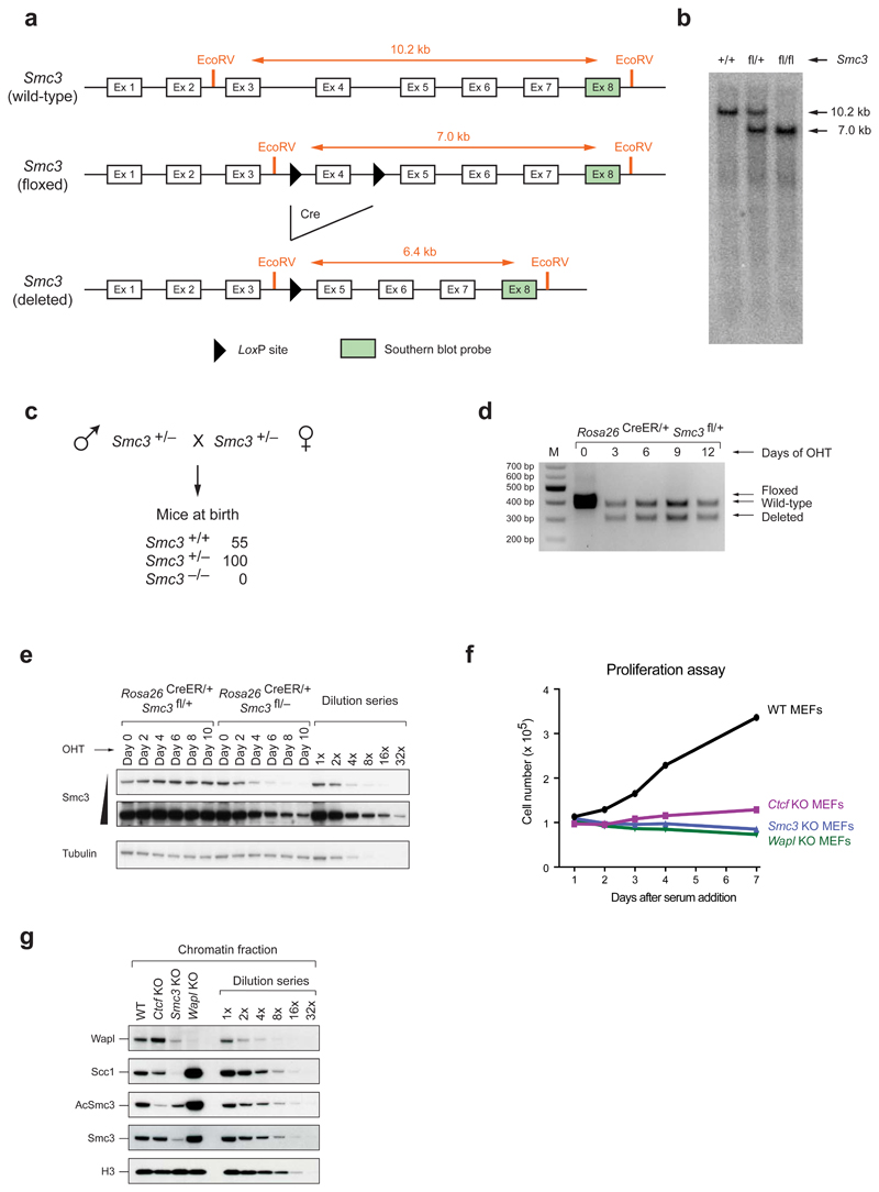
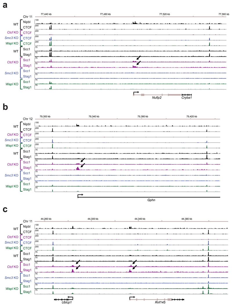
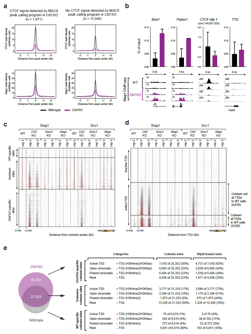
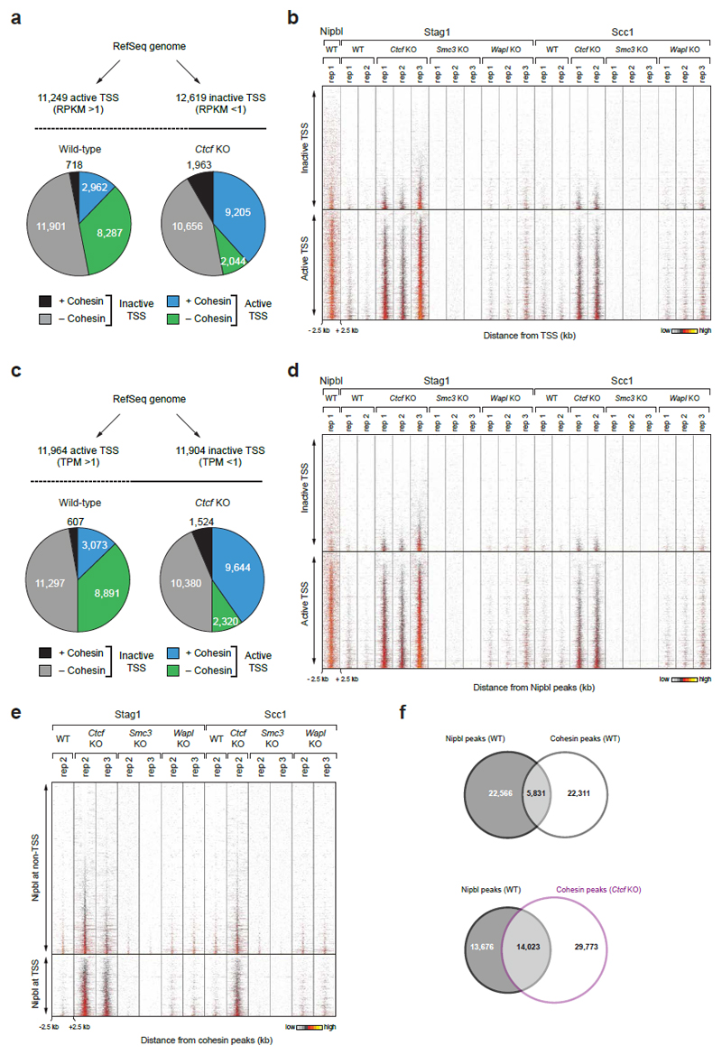
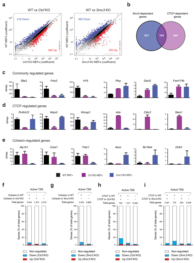
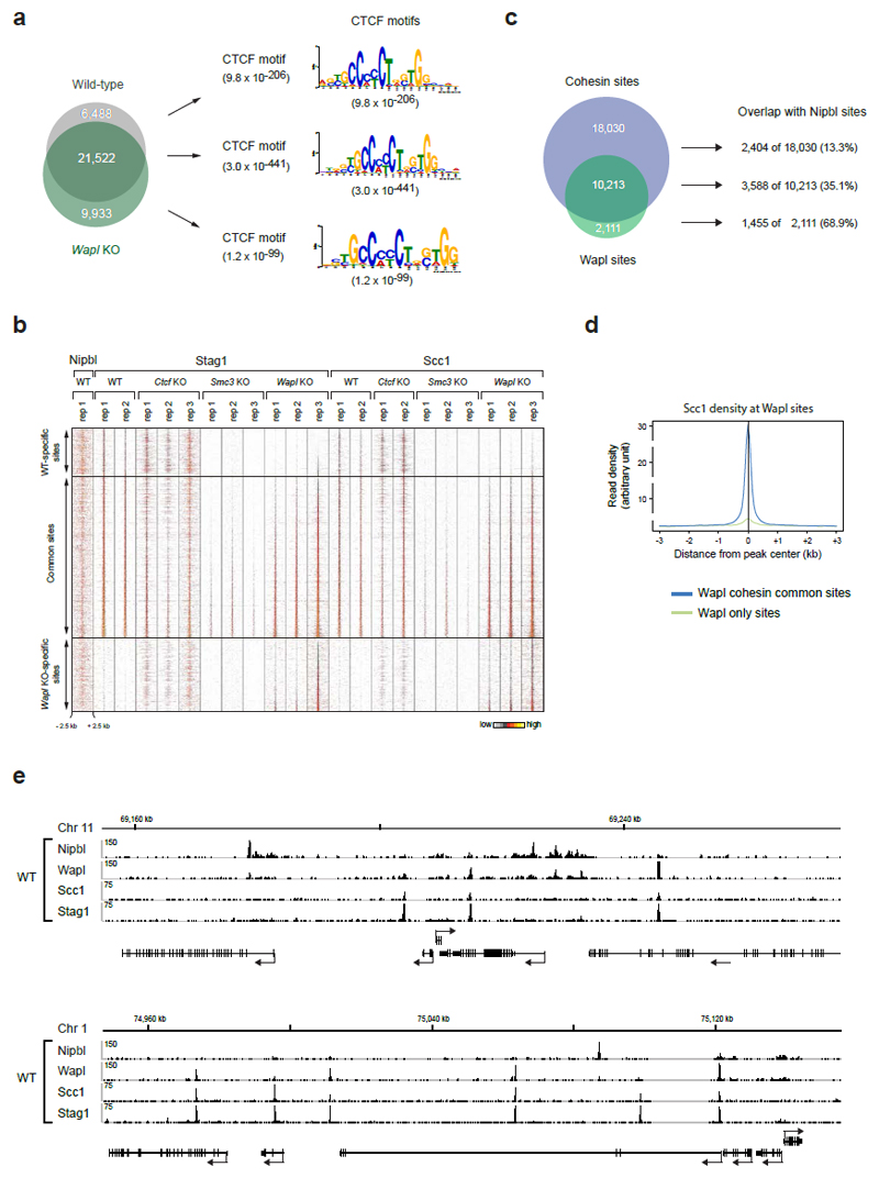
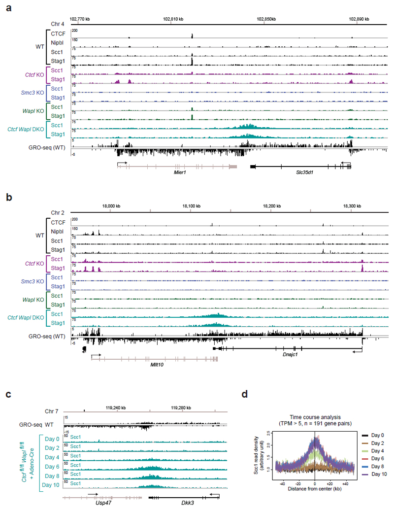
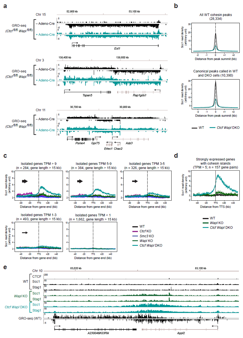
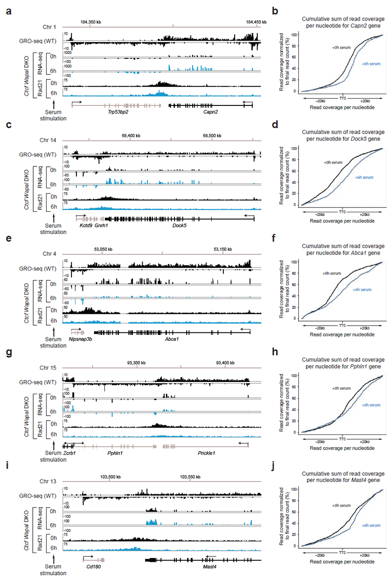
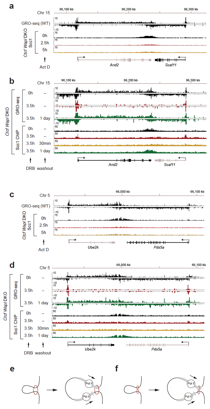
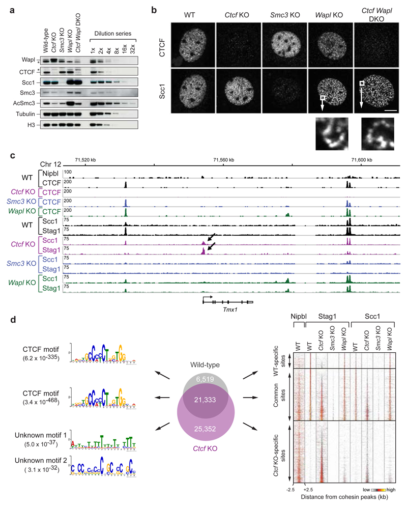
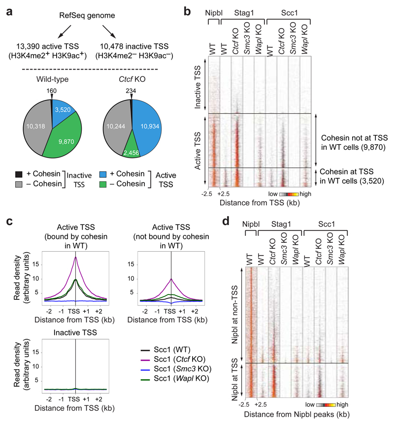
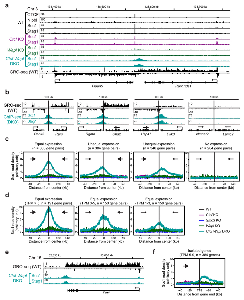
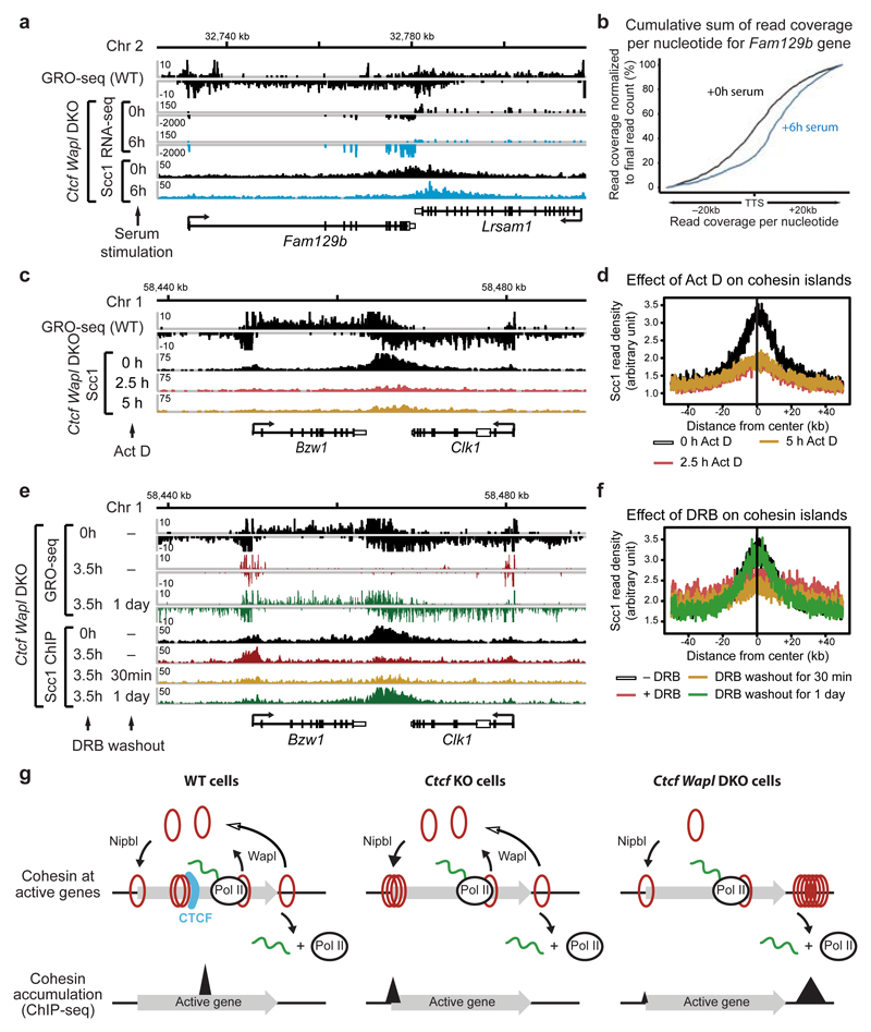
Mammalian genomes are spatially organized by CCCTC-binding factor (CTCF) and cohesin into chromatin loops and topologically associated domains, which have important roles in gene regulation and recombination. By binding to specific sequences, CTCF defines contact points for cohesin-mediated long-range chromosomal cis-interactions. Cohesin is also present at these sites, but has been proposed to be loaded onto DNA elsewhere and to extrude chromatin loops until it encounters CTCF bound to DNA. How cohesin is recruited to CTCF sites, according to this or other models, is unknown. Here we show that the distribution of cohesin in the mouse genome depends on transcription, CTCF and the cohesin release factor Wings apart-like (Wapl). In CTCF-depleted fibroblasts, cohesin cannot be properly recruited to CTCF sites but instead accumulates at transcription start sites of active genes, where the cohesin-loading complex is located. In the absence of both CTCF and Wapl, cohesin accumulates in up to 70 kilobase-long regions at 3'-ends of active genes, in particular if these converge on each other. Changing gene expression modulates the position of these 'cohesin islands'. These findings indicate that transcription can relocate mammalian cohesin over long distances on DNA, as previously reported for yeast cohesin, that this translocation contributes to positioning cohesin at CTCF sites, and that active genes can be freed from cohesin either by transcription-mediated translocation or by Wapl-mediated release.


Conflict of interest statement

Competing Financial Interests

The authors declare no competing financial interests.

Figures

Extended Data Figure 1
Extended Data Figure 1. Characterization of conditional Smc3 knockout cells.
a, Schematic representation of the wild-type, floxed (fl) and deleted (Δ) Smc3 alleles (after elimination of the neomycin resistance gene). EcoRV fragments, which were used for allele identification by Southern blot analysis with the indicated exon 8 probe, are shown together with their length (in kb). b, Southern blot analysis of tail DNA from wild-type, Smc3fl/+ and Smc3fl/fl mice. c, Absence of Smc3–/– offspring at birth. The genotype of newborn mice from intercrosses of Smc3+/– mice was determined by PCR genotyping. d, Deletion of the floxed Smc3 allele was detected by PCR genotyping in primary Rosa26CreER/+ Smc3fl/+ MEFs at the indicated days after 4-hydroxytamoxifen (OHT) addition. e, The level of Smc3 protein depletion in primary Rosa26CreER/+ Smc3fl/– MEFs was analyzed every second day after OHT addition by immunoblot analysis of whole cell extracts. Control Rosa26CreER/+ Smc3fl/+ MEFs were additionally analyzed together with a dilution series of the day-0 sample. A long and short exposure of the Smc3 immunoblot is shown. f, The efficiency of protein depletion was analyzed by immunoblot analysis of chromatin extracts from starved MEFs at day 10 after OHT treatment (Smc3 and Wapl KO cells) or Adeno-Cre virus infection (Ctcf KO cells). The wild-type chromatin sample was diluted up to 1:32 in order to estimate the relative reduction in protein levels (CTCF: > 4x, Wapl: > 8x and Smc3: > 16x). g, Proliferation capacity of WT MEFs and Ctcf, Smc3 and Wapl KO MEFs. The indicated serum-starved cells were stimulated with 10% fetal calf serum, and cell numbers were measured every day using the Casy counter. All three KO cells failed to respond to proliferate in contrast to wild-type MEFs.
Extended Data Figure 2
Extended Data Figure 2. Cohesin relocation in Ctcf KO MEFs.
Binding of Nipbl, CTCF and cohesin (Stag1 and Scc1) at the Nufip2 (a), Gphn (b) and Ublcp1/Rnf145 (c) genes was determined by ChIP-seq analysis in WT MEFs and Ctcf, Smc3 and Wapl KO MEFs.
Extended Data Figure 3
Extended Data Figure 3. Analysis of Ctcf KO-specific cohesin sites.
a, Density profiles of CTCF and Stag1 binding at cohesin sites that are commonly found in WT and Ctcf KO cells. The cohesin sites were subdivided based on the detection or absence of a CTCF peak in Ctcf KO cells as determined by the MACS peak-calling program. b, The enrichment of Stag1 binding (n=2) relative to input is shown for WT (black) and Ctcf KO cells (purple) at two TSSs, one CTCF sites and one transcription termination site (TTS). Two independent biological experiments were performed and normalized values with the corresponding standard deviations were plotted. c-d, Cohesin (Stag1 and Scc1) ChIP-seq data of replica (rep) experiments are shown as density heat maps for (c) active and inactive TSSs and (d) TSS- and non-TSS-associated Nipbl-binding sites. Binding data are shown for a region extending from -2.5 kb to +2.5 kb relative to the cohesin peak summit. Heat maps were sorted according to the density of Stag1 binding in Ctcf KO cells (rep 2). A density scale from low (grey) to high (yellow) is shown. e, Categorization of WT-specific, Ctcf KO-specific and commonly found cohesin sites according to their location at active TSSs (H3K4me2+ H3K9ac+) or non-TSS regions with open chromatin (H3K4me2+ H3K9ac+), poised chromatin (H3K4me2+) and no active chromatin marks (rest). Note that one TSS can bind several cohesin sites. Therefore the number of cohesin-bound TSSs and TSS-bound cohesin sites are not necessarily identical.
Extended Data Figure 4
Extended Data Figure 4. Cohesin redistribution to transcriptional start sites in Ctcf KO MEFs.
a, Binding of cohesin at transcription start sites (TSSs) of active and inactive genes, which were defined by RNA-seq analysis. Genes with an RPKM > 1 were considered as active, whereas genes with an RPKM < 1 were classified as inactive. Pie charts indicate the relative binding of cohesin at all annotated TSSs of the RefSeq genome (mm9) in WT and Ctcf KO MEFs. b, Density heat map of cohesin and Nipbl binding at active and inactive TSSs as defined in a. Active and inactive TSSs were sorted according to the read density of Stag1 binding in Ctcf KO cells (replica 3). c, Binding of cohesin at TSSs of active and inactive genes, which were defined by GRO-seq analysis. Genes with a TPM > 1 were considered as active, whereas genes with an TPM < 1 were classified as inactive. Pie charts indicate the relative binding of cohesin at all annotated TSSs of the RefSeq genome (mm9) in WT and Ctcf KO cells. d, Density heat map of cohesin and Nipbl binding at active and inactive TSSs as defined in c. Active and inactive TSSs were sorted according to the read density of Stag1 binding in Ctcf KO (replica 3). A density scale from low (grey) to high (yellow) is shown (b,d). e, Heat map of Nipbl and cohesin binding at Nipbl peaks in MEFs of the indicated genotypes. The Nipbl peaks were subdivided according to their TSS localization. Peaks were sorted according to the Stag1 binding density in Ctcf KO cells (replica 3). f, Venn diagram indicating the overlap between Nipbl and cohesin peaks in WT or Ctcf KO MEFs.
Extended Data Figure 5
Extended Data Figure 5. Identification of CTCF- and cohesin-regulated genes.
a, Scatter plot of gene expression differences between WT and Ctcf KO or Smc3 KO MEFs, based on 2 and 4 independent RNA-seq experiments, respectively. The normalized expression data of individual genes in the two cell types are plotted as coefficient value. Each symbol represents one gene. Genes with an expression difference of > 2-fold, an adjusted P value of < 0.1 and an RPKM value of > 1 in WT or KO cells are colored in blue or red, corresponding to down- or up-regulated genes in the indicated KO MEFs, respectively. For evaluation of the RNA-seq data, see Online Methods. b, Overlap between CTCF- and cohesin-regulated genes, shown as a Venn diagram. c-e, Expression of selected regulated genes in WT (black), Ctcf KO (purple) and Smc3 KO (blue) MEFs. The expression of genes, which are commonly regulated by CTCF and cohesin (c), by CTCF alone (d) or by cohesin alone (e), is shown as normalized expression value (RPKM) based on 10 (WT), 2 (Ctcf KO) or 4 (Smc3 KO) independent RNA-seq experiments. RPKM, reads per kilobase of exon per million mapped sequence reads. f,h, Minimal correlation between CTCF-dependent gene regulation and cohesin (f) or CTCF (h) binding at active promoters. Genes that were down- or up-regulated in Ctcf KO MEFs are shown as percentage of all genes present in the three indicated gene groups that were defined by the presence or absence of cohesin binding at active TSSs in WT and/or Ctcf KO cells. g,i, Little correlation between Smc3-dependent gene regulation and cohesin (g) or CTCF (i) binding at active promoters. Genes that were down- or up-regulated in Smc3 KO MEFs are shown as percentage of all genes that were defined by the presence or absence of cohesin binding at active TSSs in WT cells.
Extended Data Figure 6
Extended Data Figure 6. Genomic localization of cohesin in Wapl KO cells.
a. The cohesin-binding sites detected in wild-type and Wapl KO cells were subdivided into WT-specific, Wapl KO-specific and common peaks, as indicated by the Venn diagram. For each subgroup, the most significant DNA-binding motif is shown. The different motifs were detected with the E-value indicated in brackets. b. Heat maps of Nipbl and cohesin binding for the different subgroups. Cohesin peaks were blotted on the vertical axis for a region extending from -2.5 kb to +2.5 kb relative to the cohesin peak summit (horizontal axis) and were sorted in each subgroup according to the density of Stag1 binding in Wapl KO cells (replica 3). A density scale from low (grey) to high (yellow) is shown. c, Venn diagram of Wapl and cohesin peaks in WT MEFs. The individual subgroups were further overlapped with Nipbl sites. d, Density profiles of Scc1 binding are shown for Wapl only sites (green) and cohesin/Wapl common sites (blue). Note that cohesin is also enriched (even though at low level, which is not detected by the peak calling algorithm we used) at "Wapl only" sites, consistent with our observation that the association of Wapl with chromatin depends on cohesin and Extended Data 1f). e, Examples of the distribution of Nipbl, Wapl, and cohesin (Stag1 and Scc1) at two genomic regions, one on chromosome 11 and the other on chromosome 1, as determined by ChIP-seq analysis in WT MEFs.
Extended Data Figure 7
Extended Data Figure 7. Cohesin islands in Ctcf Wapl DKO MEFs.
Binding of CTCF, Nipbl and cohesin (Stag1 and Scc1) at the convergently transcribed genes Mier1 and Slc35d1 (a) and Mllt1 and Dnajc1 (b) was determined by ChIP-seq analysis in WT MEFs, Ctcf, Smc3 and Wapl KO MEFs and Ctcf Wapl DKO MEFs. c,d. Time course analysis of the appearance of cohesin islands upon Ctcf and Wapl deletion in Adeno-Cre infected MEFs. c, Scc1 accumulation in the 3’ region of the convergently transcribed Usp47 and Dkk3 genes at the indicated days after Adeno-Cre infection. d, Density profiles of Scc1 accumulation at convergently transcribed genes in response to Adeno-Cre infection. Scc1 binding was centered in the middle of the intervening region between gene pairs with a similarly strong transcription activity (TPM > 5).
Extended Data Figure 8
Extended Data Figure 8. Cohesin islands at isolated genes depend on gene transcription.
a, Similar transcriptional activity of individual genes in WT and Ctcf Wapl DKO MEFs. GRO-seq data are shown for three different gene regions in Ctcf fl/fl Wapl fl/fl MEFs before and after Adeno-Cre infection. b. Density profiles of Scc1 accumulation in wild-type and Ctcf Wapl DKO cells at all wild-type cohesin peaks (28,334) or at overlapping canonical peaks (10,390) identified by peak calling in WT and Ctcf Wapl DKO cells. c, The transcriptional activity determines the amount of cohesin accumulation in the 3’ region of isolated genes lacking a neighboring downstream gene. Density profiles of Scc1 binding are shown for 5 groups of genes with decreasing GRO-seq signals (TPM value of > 9, 5-9, 3-5, 1-3 and < 1). d, For better visualization of cohesin islands in Wapl KO cells, we restricted our analysis on genes that were > 15kb in length and highly expressed (TPM > 5). The genes were further filtered based on the presence of a cohesin island in Ctcf Wapl KO cells and based on the absence of any intragenic CTCF site (assuming that a CTCF site might prevent proper cohesin pushing along the DNA). Density profiles of Scc1 in wild-type (WT, black), Wapl KO (green) and Ctcf Wapl KO (turquoise) cells are shown. e, Examples of cohesin islands formation in Wapl KO cells is shown for the A230046K03Rik/Appl2 locus.
Extended Data Figure 9
Extended Data Figure 9. Transcriptional changes induced by serum stimulation affect positioning of cohesin islands.
a,c,e,g,i. Binding of Scc1 and mRNA profiling upon serum stimulation (20% fetal bovine serum) at five genomic regions. The observed differences in cohesin island positioning correlate with increased or decreased mRNA levels of the respective genes as measured by their RPKM value (a, Capn2: 104.88 → 248.32; c, Dock5: 4.89 → 14.06; e, Abca1: 11.51 → 2.27; g, Pphln1: 5.13 → 8.00 and Prickle1: 14.19 → 7.86; i, Mast4: 7.70 →16.68). b,d,f.h,j Visualization of the altered shape of cohesin islands by plotting the cumulative sum of reads per nucleotide starting from -20 kb to + 20 kb after the transcription termination sites of Capn2 (b), Dock5 (d), Abca1 (f), Pphln1 (h) and Mast4 (j) gene (see Online Methods).
Extended Data Figure 10
Extended Data Figure 10. Transcriptional inhibition and disappearance of cohesin islands.
a,c. Loss of cohesin islands in response to transcriptional inhibition by actinomycin D (Act D; 5 μg/ml) for 2.5 and 5 h followed by Scc1 profiling in Ctcf Wapl DKO cells. b,d. Dis- and reapperance of cohesin islands in response to inhibition of RNA polymerase II elongation by 5,6-dichlorobenzimidazole 1-β-D-ribofuranoside (DRB; 100 μM) and its subsequent removal in Ctcf Wapl DKO cells, respectively. The same convergently transcribed gene pairs are shown for the Act D and DRB experiments (a,b Arid2 and Scaf11 and c,d Ube2k and Pds5a). Please note that Nipbl localization in Drosophila is not affected by transcriptional inhibitors, implying that Nipbl-cohesin interactions may not be sufficient to explain cohesin accumulation at TSSs in DRB treated cells. e,f, Speculative models of how transcription could move one (e) or two (f) cohesin rings to mediate loop extrusion.
Figure 1
Figure 1. Cohesin distribution in wild-type, Ctcf, Smc3 and Wapl knockout MEFs.
a, Immunoblot analysis of whole cell extracts of quiescent knockout MEFs including dilution series of wild-type sample. b, Fluorescence microscopy with Scc1 and CTCF antibodies. Size bar, 10 μm. Below: higher magnification of Scc1 staining. c, Binding of CTCF, Nipbl, Stag1 and Scc1 at the Tmx1 locus, as determined by ChIP-seq. d, Analysis of cohesin-binding site distribution in wild-type and Ctcf KO cells (Venn diagram). Left: DNA-binding motif prediction with indicated E-value. Right: heat maps of cohesin and Nipbl binding in different KO cells (sorted according to Stag1 binding in Ctcf KO cells).
Figure 2
Figure 2. Cohesin redistribution to transcriptional start sites in Ctcf KO MEFs.
a, Cohesin binding at active (H3K4me2+ H3K9ac+) and inactive (H3K4me2 H3K9ac) TSSs. Pie charts indicate cohesin binding at all annotated TSSs in wild-type and Ctcf KO cells. b, Density heat map of Stag1, Scc1 and Nipbl binding at active and inactive TSSs, data sorted by Stag1 binding in Ctcf KO MEFs. Active TSSs were subdivided based on cohesin binding in wild-type cells (right). c, Density profiles of Scc1 binding at active TSSs, subdivided as in b, and at inactive TSSs in MEFs of the indicated genotypes. d, Density heat map of Nipbl, Stag1 and Scc1 binding at Nipbl sites, which are grouped by TSS localization. Reads sorted according to Stag1 binding in Ctcf KO cells.
Figure 3
Figure 3. Cohesin accumulation at sites of convergent transcription in Ctcf Wapl DKO MEFs.
a, Cohesin accumulation in the 3’ region of Tspan5 and Rap1gds1. Binding of CTCF, Nipbl, Stag1 and Scc1 was determined by ChIP-seq, and the transcriptional activity was measured by GRO-seq in wild-type MEFs. b, c, Cohesin island formation in Ctcf Wapl DKO cells at convergent gene pairs with different transcriptional activity. Individual example (b) and Scc1 density plots (c) for each category is shown. d, Density profiles of Scc1 binding for equally transcribed gene pairs with different transcriptional activity (TPM > 5, 3-5 and 1-3). e, Presence of cohesin islands at the actively transcribed isolated Ext1 gene. f, Scc1-binding profile for all isolated transcribed genes (TPM 5-9).
Figure 4
Figure 4. Transcription is required for the generation of cohesin islands.
a, Binding of Scc1 and mRNA profiling in DKO cells upon serum stimulation at the Fam129b/Lrsam1 gene pair. b, Shift in cohesin island positioning is visualized by the cumulative sum of reads per nucleotide (see Online Methods). c, Loss of cohesin island at the Bzw1/Clk1 gene pair upon transcriptional inhibition by actinomycin D in DKO cells. d. Density profiles of Scc1 accumulation at convergently transcribed genes in response to actinomycin D treatment. e. Dis- and re-apperance of a cohesin island upon temporal inhibition of RNA polymerase II by DRB at the Bzw1/Clk1 gene pair in DKO cells. f. Density profiles of Scc1 accumulation at convergently transcribed genes in response to DRB treatment. g, Model of cohesin movement and accumulation at transcribed genes. Cohesin rings are indicated by red circles and cohesin peaks as determined by ChIP-seq by black triangles.

Similar articles

Cited by

References

    1. Hadjur S, et al. Cohesins form chromosomal cis-interactions at the developmentally regulated IFNG locus. Nature. 2009;460:410–413. - PMC - PubMed
    1. Nativio R, et al. Cohesin is required for higher-order chromatin conformation at the imprinted IGF2-H19 locus. PLoS Genet. 2009;5:e1000739. - PMC - PubMed
    1. Dixon JR, et al. Topological domains in mammalian genomes identified by analysis of chromatin interactions. Nature. 2012;485:376–380. - PMC - PubMed
    1. Zuin J, et al. Cohesin and CTCF differentially affect chromatin architecture and gene expression in human cells. Proc Natl Acad Sci USA. 2014;111:996–1001. - PMC - PubMed
    1. Sofueva S, et al. Cohesin-mediated interactions organize chromosomal domain architecture. EMBO J. 2013;32:3119–3129. - PMC - PubMed

Publication types

MeSH terms