

Recent Advances in Bioink Design for 3D Bioprinting of Tissues and Organs
- PMID: 28424770
- PMCID: PMC5380738
- DOI: 10.3389/fbioe.2017.00023
Recent Advances in Bioink Design for 3D Bioprinting of Tissues and Organs
Abstract
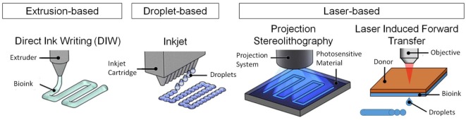
There is a growing demand for alternative fabrication approaches to develop tissues and organs as conventional techniques are not capable of fabricating constructs with required structural, mechanical, and biological complexity. 3D bioprinting offers great potential to fabricate highly complex constructs with precise control of structure, mechanics, and biological matter [i.e., cells and extracellular matrix (ECM) components]. 3D bioprinting is an additive manufacturing approach that utilizes a "bioink" to fabricate devices and scaffolds in a layer-by-layer manner. 3D bioprinting allows printing of a cell suspension into a tissue construct with or without a scaffold support. The most common bioinks are cell-laden hydrogels, decellulerized ECM-based solutions, and cell suspensions. In this mini review, a brief description and comparison of the bioprinting methods, including extrusion-based, droplet-based, and laser-based bioprinting, with particular focus on bioink design requirements are presented. We also present the current state of the art in bioink design including the challenges and future directions.
Keywords: additive manufacturing; biofabrication; cell printing; extracellular matrix; hydrogel; regenerative medicine; tissue engineering.
Figures

Similar articles
-
Bioprinting 101: Design, Fabrication, and Evaluation of Cell-Laden 3D Bioprinted Scaffolds.Tissue Eng Part A. 2020 Mar;26(5-6):318-338. doi: 10.1089/ten.TEA.2019.0298. Tissue Eng Part A. 2020. PMID: 32079490 Free PMC article.
-
Advancing bioinks for 3D bioprinting using reactive fillers: A review.Acta Biomater. 2020 Sep 1;113:1-22. doi: 10.1016/j.actbio.2020.06.040. Epub 2020 Jul 2. Acta Biomater. 2020. PMID: 32622053 Review.
-
3D Bioprinting of Human Tissues: Biofabrication, Bioinks, and Bioreactors.Int J Mol Sci. 2021 Apr 12;22(8):3971. doi: 10.3390/ijms22083971. Int J Mol Sci. 2021. PMID: 33921417 Free PMC article. Review.
-
Designing Decellularized Extracellular Matrix-Based Bioinks for 3D Bioprinting.Adv Healthc Mater. 2020 Dec;9(24):e2000734. doi: 10.1002/adhm.202000734. Epub 2020 Jul 21. Adv Healthc Mater. 2020. PMID: 32691980 Review.
-
Recent Trends in Decellularized Extracellular Matrix Bioinks for 3D Printing: An Updated Review.Int J Mol Sci. 2019 Sep 18;20(18):4628. doi: 10.3390/ijms20184628. Int J Mol Sci. 2019. PMID: 31540457 Free PMC article. Review.
Cited by
-
Fused Filament Fabrication 3D Printing Parameters Affecting the Translucency of Polylactic Acid Parts.Polymers (Basel). 2024 Oct 10;16(20):2862. doi: 10.3390/polym16202862. Polymers (Basel). 2024. PMID: 39458689 Free PMC article.
-
Advanced 4D Bioprinting Technologies for Brain Tissue Modeling and Study.Int J Smart Nano Mater. 2019;10(3):177-204. doi: 10.1080/19475411.2019.1631899. Epub 2019 Jul 3. Int J Smart Nano Mater. 2019. PMID: 32864037 Free PMC article.
-
Formation of cell spheroids using Standing Surface Acoustic Wave (SSAW).Int J Bioprint. 2017 Jan 17;4(1):130. doi: 10.18063/IJB.v4i1.130. eCollection 2018. Int J Bioprint. 2017. PMID: 33102912 Free PMC article.
-
Translational Application of Microfluidics and Bioprinting for Stem Cell-Based Cartilage Repair.Stem Cells Int. 2018 Feb 20;2018:6594841. doi: 10.1155/2018/6594841. eCollection 2018. Stem Cells Int. 2018. PMID: 29535776 Free PMC article. Review.
-
The sculpting tool in bioprinting: research and application progress of sacrificial inks.Front Bioeng Biotechnol. 2025 Jun 25;13:1486459. doi: 10.3389/fbioe.2025.1486459. eCollection 2025. Front Bioeng Biotechnol. 2025. PMID: 40635697 Free PMC article. Review.
References
-
- Barron J. A., Spargo B. J., Ringeisen B. R. (2004a). Biological laser printing of three dimensional cellular structures. Appl. Phys. A Mater. Sci. Process. 79, 1027–1030.10.1007/s00339-004-2620-3 - DOI
Publication types
LinkOut - more resources
Full Text Sources
Other Literature Sources