. 2017:2017:2023853.
doi: 10.1155/2017/2023853. Epub 2017 Mar 23.

Postembedding Decalcification of Mineralized Tissue Sections Preserves the Integrity of Implanted Biomaterials and Minimizes Number of Experimental Animals

Affiliations

Postembedding Decalcification of Mineralized Tissue Sections Preserves the Integrity of Implanted Biomaterials and Minimizes Number of Experimental Animals

Thaqif El Khassawna et al. Biomed Res Int. 2017.

Abstract

Bone histology of decalcified or undecalcified samples depends on the investigation. However, in research each method provides different information to answer the scientific question. Decalcification is the first step after sample fixation and governs what analysis is later feasible on the sections. Besides, decalcification is favored for immunostaining and in situ hybridization. Otherwise, sample decalcification can be damaging to bone biomaterials implants that contains calcium or strontium. On the other hand, after decalcification mineralization cannot be assessed using histology or imaging mass spectrometry. The current study provides a solution to the hardship caused by material presence within the bone tissue. The protocol presents a possibility of gaining sequential and alternating decalcified and undecalcified sections from the same bone sample. In this manner, investigations using histology, protein signaling, in situ hybridization, and mass spectrometry on the same sample can better answer the intended research question. Indeed, decalcification of sections and grindings resulted in well-preserved sample and biomaterials integrity. Immunostaining was comparable to that of classically decalcified samples. The study offers a novel approach that incites correlative analysis on the same sample and reduces the number of processed samples whether clinical biopsies or experimental animals.


Figures

Figure 1
Figure 1
A schematic explains the experimental design and the route of samples according to the desired stain, calcification status, and embedding medium.
Figure 2
Figure 2
Specific osteoclast staining after postembedding decalcification of rat femur. Comparison of undecalcified bone sections with decalcified bone reflects no alteration of bone integrity. (a–c) Preembedding decalcified and paraffin embedded bone samples show TRAP positive cells. (d–f) Undecalcified PMMA section shows the specificity of TRAP staining. (g–i) Postembedding decalcification result is comparable to both decalcified and undecalcified sections. (c, f, and i) Magnified images show the multinucleated osteoclasts on bone surface. Images (a), (d), and (g) were acquired by 5x magnification objective; then individual tiles were stitched together by Leica application suite (LASX) software. Images (b), (e), and (h) were acquired by 40x magnification objective. Images (c), (f), and (i) were acquired by 100x magnification objective.
Figure 3
Figure 3
Postembedding decalcification of rat femur preserves antigen specificity in bone tissue. (a–c) Preembedding decalcified bone samples embedded in paraffin show positive ColI signaling in bone matrix. (d–f) PMMA undecalcified section shows the specificity of ColI immunostaining. (g–i) Postembedding decalcification shows no discrepancies in signal intensity and localization. Images (a), (d), and (g) were acquired by 5x magnification objective; then individual tiles were stitched together by Leica application suite (LASX) software. Images (b), (e), and (h) were acquired by 5x magnification objective. Images (c), (f), and (i) were acquired by 10x magnification objective.
Figure 4
Figure 4
Postembedding decalcification of rat femur preserves the integrity of calcium-containing materials and allows investigating osteocytes. The SrCPC cement in the fracture defect was embedded in PMMA T9100. (a-b) von Kossa/van Gieson staining of undecalcified bone sample differentiates soft tissue around the fracture defect (red) and stains ossified tissue and materials in black. (c-d) Silver staining of postembedding decalcified sections visualizes osteocytes within the matrix and differentiates the implant from the surrounding tissue. Images (a) and (c) were acquired by 5x magnification objective; then individual tiles were stitched together by Leica application suite (LASX) software. Images (b) and (d) were acquired by 40x magnification objective while inset of image (d) was acquired by 100x magnification objective.
Figure 5
Figure 5
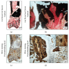
Histological stains of undecalcified tibia with intramedullary pin aligned in compliance with decalcified bone. The bone samples were embedded in PMMA T7200. Masson-Goldner staining of undecalcified bone shows the presence of soft tissue around the implant region and the magnified image shows the clear view of osteoid (a–c). The decalcified bone section shows the comparative view and presence of soft tissue around the implant region (d–f). The magnified image shows the view of osteocyte canaliculi. Images (a) and (d) were acquired by 5x magnification objective; then individual tiles were stitched together by Leica application suite (LASX) software. Images (b) and (e) were acquired by 40x magnification objective. Images (c) and (f) were acquired by 100x magnification objective.
Figure 6
Figure 6
Silver stain visualizes the completely decalcified biomaterial and the osteocytes. Undecalcified femur with SrCPC in PMMA T9100 sections shows the clear portions of minerals and organic compounds. (a) Ca+ distribution accounts for CPC and mineralized bone matrix. (b) Strict presence of Sr+ within the SrCPC area and its vicinity. (c) Organic compound at the bone matrix areas and not the CPC area. (d) Overlay of minerals (Ca+: green; Sr+: blue; and organic compound: red) serves as a map of portions distribution before decalcification. (e-f) Collagenous portion before (e) decalcification in the same areas as after (f) decalcification. (g) The Ag+ staining corresponds to the areas of organic compounds. (h) Overlay of Ca+ (red) before decalcification and Ag+ after decalcification (green) hints that the Ag+ is visible after mineral removal. (i-j) Light microscopic imaging of decalcified bone section shows the comparative view of the implant and the surrounding tissue. Furthermore, osteocytes and cell canaliculi are evident in the bone matrix. Image (i) was acquired by 5x magnification objective; then individual tiles were stitched together by Leica application suite (LASX) software. (j) was acquired by 40x magnification objective whereas inset was acquired by 100x magnification objective.
Figure 7
Figure 7
Chemical investigations show preserved collagenous portion before and after decalcification. (a) Complete depletion of mineral constituents within tissue and material. (b) Minerals masked the signal of organic compounds before decalcification; however, after decalcification the surface analysis depicts demineralized areas as cartilaginous portions. The overlay shows that portions before decalcification were highly preserved.

Similar articles

Cited by

References

    1. Goldhahn J., Suhm N., Goldhahn S., Blauth M., Hanson B. Influence of osteoporosis on fracture fixation—a systematic literature review. Osteoporosis International. 2008;19(6):761–772. doi: 10.1007/s00198-007-0515-9. - DOI - PubMed
    1. Low K. L., Tan S. H., Zein S. H. S., Roether J. A., Mouriño V., Boccaccini A. R. Calcium phosphate-based composites as injectable bone substitute materials. Journal of Biomedical Materials Research. Part B Applied Biomaterials. 2010;94(1):273–286. doi: 10.1002/jbm.b.31619. - DOI - PubMed
    1. Ginebra M.-P., Traykova T., Planell J. A. Calcium phosphate cements: competitive drug carriers for the musculoskeletal system? Biomaterials. 2006;27(10):2171–2177. doi: 10.1016/j.biomaterials.2005.11.023. - DOI - PubMed
    1. Gong T., Wang Z., Zhang Y., et al. Preparation, characterization, release kinetics, and in vitro cytotoxicity of calcium silicate cement as a risedronate delivery system. Journal of Biomedical Materials Research. Part A. 2014;102(7):2295–2304. doi: 10.1002/jbm.a.34908. - DOI - PubMed
    1. Wu C.-C., Wang C.-C., Lu D.-H., Hsu L.-H., Yang K.-C., Lin F.-H. Calcium phosphate cement delivering zoledronate decreases bone turnover rate and restores bone architecture in ovariectomized rats. Biomedical Materials. 2012;7(3) doi: 10.1088/1748-6041/7/3/035009.035009 - DOI - PubMed

LinkOut - more resources