. 2017 May;22(3):429-443.
doi: 10.1007/s12192-017-0795-8. Epub 2017 Apr 19.

Oxidative protein modification alters proteostasis under acute hypobaric hypoxia in skeletal muscles: a comprehensive in vivo study

Affiliations

Oxidative protein modification alters proteostasis under acute hypobaric hypoxia in skeletal muscles: a comprehensive in vivo study

Akanksha Agrawal et al. Cell Stress Chaperones. 2017 May.

Abstract

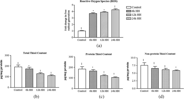
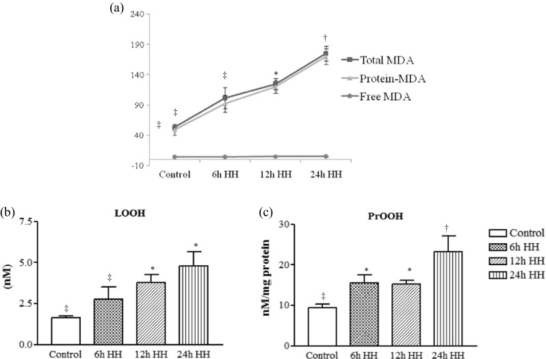
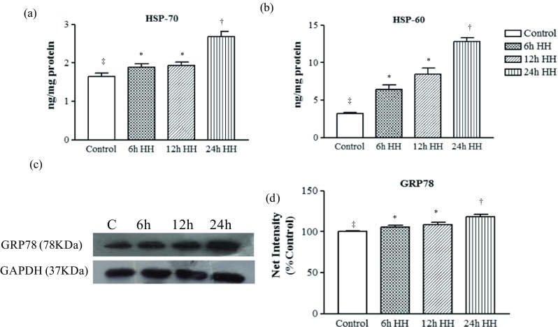
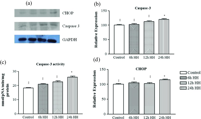
While numerous maladies are associated with hypobaric hypoxia, muscle protein loss is an important under studied topic. Hence, the present study was designed to investigate the mechanism of muscle protein loss at HH. SD rats were divided into normoxic rats, while remaining rats were exposed to simulated hypoxia equivalent to 282-torr pressure (equal to an altitude of 7620 m, 8% oxygen), at 25 °C for 6, 12, and 24 h. Post-exposure rats were sacrificed and analysis was performed. Ergo, muscle loss-related changes were observed at 12 and 24 h post-HH exposure. An increased reactive oxygen species production and decreased thiol content was observed in HH-exposed rats. This disturbance caused substantial protein oxidative modification in the form of protein carbonyl content and advanced oxidation protein products. The analysis showed increase levels of bityrosine, oxidized tryptophan, lysine conjugate, lysine conjugate with MDA, protein hydroperoxide, and protein-MDA product. These changes were also in agreement with increase in lipid hydroperoxides and MDA content. HSP-70 and HSP-60 were upregulated significantly, and this finding is corroborated with increase in ER stress biomarker, GRP-78. Overloading of cells with misfolded proteins further activated degradative machinery. Consequently, pro-apoptotic signaling cascade, caspase-3, and C/EBP homologous protein were also activated in 24-h HH exposure. Release of tryptophan and tyrosine was also increased with 24-h HH exposure, indicated protein degradation. Elevation in resting intracellular calcium ion, [Ca2+]i, was also observed at 12- and 24-h HH exposure. The present study provides a detailed mechanistic representation of muscle protein loss during HH exposure.

Keywords: Calcium; High altitude; Hypobaric hypoxia (HH); Muscle loss, proteostasis; Protein modifications.


Figures

Fig. 1
Fig. 1
Hypobaric hypoxia induced oxidative damage via ROS generation and affects intracellular sulfhydryl content in rat muscle. a Reactive oxygen species. b Total thiol content (T-SH). c Protein thiol content (Pr-SH). d Non-protein thiol content (NPr-SH). Data represents the mean ± SE; N = 5. Different symbols, double dagger, asterisk, and dagger indicate the significant differences between experimental groups (p < 0.05), while groups with matching symbols denote no difference (p ≥ 0.05)
Fig. 2
Fig. 2
Free radical generation under hypobaric hypoxic stress resultant into protein modification and the amino acid oxidation in skeletal muscle tissue homogenate by fluorescence excitation-emission spectra. a Protein carbonylation. b Advanced oxidation protein products (AOPPs). c Formation of protein-protein cross-linkage (bityrosine formation) due to presence of free radical species shows characteristic emission spectra at 380–440 nm. d Emission spectra due to the oxidation of aromatic amino acid tryptophan under hypoxic condition, showing a shift in intensity at the peak emission wavelength. e Excitation spectra of lysine conjugates measured at 325–380 nm. f Emission spectra at 425–480 nm for the lysine conjugates with lipid peroxidation products like malondialdehyde, 4-hydroxynenal through covalent attachment designated as reactive carbonyl species (RCS)-derived protein carbonylation. Data represents the mean ± SE; N = 5. Different symbols, double dagger, asterisk, and dagger indicate the significant differences between experimental groups (p < 0.05), while groups with matching symbols denote no difference (p ≥ 0.05)
Fig. 3
Fig. 3
Determination of lipid peroxidation product, PrOOH, and LOOH by the spectophotometric method, which is based on the reaction of TBA with MDA and Fe3+ with xylenol orange (XO) under acidic conditions and the formation of XO-Fe complex absorbing at 532 nm, respectively. a Total MDA (strikethrough square), protein MDA (Pr-MDA) (strikethrough triangle), and free MDA (Fr-MDA) (strikethrough circle). b LOOH represents lipid and other hydrophobic peroxides. c PrOOH representing protein hyderoperoxides. Data represents the mean ± SE; N = 5. Different symbols, double dagger, asterisk, and dagger indicate the significant differences between experimental groups (p < 0.05), while groups with matching symbols denote no difference (p ≥ 0.05)
Fig. 4
Fig. 4
Misfolded/oxidized proteins activate the heat shock proteins and ER chaperones in response to acute hypoxic stress. Graph shows the significant increase of heat shock proteins in acute hypoxic insult in muscle tissue homogenate. a HSP70. b HSP60. c Representative western blot identifying the expression of ER chaperone (GRP-78) in cytosolic tissue extract. Loading control was normalized with reference to GAPDH. d Graph represents the semiquantitative densitometric analysis of the protein expression. The net intensity was analyzed by using the ImageJ software. Data represents the mean ± SE; N = 5. Different symbols, double dagger, asterisk, and dagger indicate the significant differences between experimental groups (p < 0.05), while groups with matching symbols denote no difference (p ≥ 0.05)
Fig. 5
Fig. 5
Presence of oxidized proteins in the cell activates several degradative machinery, release of aromatic amino acids after degradation, and elevation of intracellular [Ca2+]i in muscle. a The 20S proteasome activity was analyzed by using fluorogenic substrate Succ-LLVY-AMC. b Calcium ion-dependent calpain activity was also analyzed fluorometrically by using substrate SLY-AMC. c, d Hypobaric hypoxia causes elevation of release of aromatic amino acids tyrosine and tryptophan residues after the degradation through proteasome and calpain activity. e Resting intracellular [Ca2+]i in muscle. Data represents the mean ± SE; N = 5. Different symbols, double dagger, asterisk, and dagger indicate the significant differences between experimental groups (p < 0.05), while groups with matching symbols denote no difference (p ≥ 0.05)
Fig. 6
Fig. 6
Acute hypoxic stress led to activation of pro-apoptotic markers. a Representative western blot identifying the expression of apoptotic marker of ER stress, caspase-3, and CHOP. Loading control was normalized with reference to GAPDH. b Graph represents densitometric analysis of the protein expression of caspase 3 using ImageJ software. c Caspase 3 activity. d Graph represents the densitometric analysis of CHOP using ImageJ software. Data represents the mean ± SE; N = 5. Different symbols, double dagger, asterisk, and dagger indicate the significant differences between experimental groups (p < 0.05), while groups with matching symbols denote no difference (p ≥ 0.05)
Fig. 7
Fig. 7
Myocyte architecture undergoes changes under hypobaric hypoxia exposure. Light micrographs showing hematoxylin-eosin staining of muscle; arrows indicate the area of maximal damage. a Control muscle cut in cross section has normal morphology with peripheral nuclei and abundant myofibrils within individual muscle fibers evenly separated by the extracellular space. b The 6-h HH-exposed rat muscle showing smaller in size with more space between myocytes (indicated by black arrow). c The 12-h HH-exposed rat muscle showing more space between myocytes; along with this, irregularity was also observed in myocytes (indicated by left-pointing white arrow). d Rat muscle exposed with 24-h HH exposure illustrated degeneration of muscle fibers (indicated by white triangle). The scale bar at the bottom represents 10 μm (ad)
Fig. 8
Fig. 8
Diagrammatic representation of mechanism approach to elucidate the effect of hypobaric hypoxia-induced muscle loss

References

    1. Alderton JM, Steinhardt RA. Calcium influx through calcium leak channels is responsible for the elevated levels of calcium-dependent proteolysis in dystrophic myotubes. J Biol Chem. 2000;275:9452–9460. doi: 10.1074/jbc.275.13.9452. - DOI - PubMed
    1. Baehr LM, West DWD, Marcotte G, Marshall AG, De Sousa LG, Baar K, Bodine SC. Age related deficits in skeletal muscle recovery following disuse are associated with neuromuscular junction instability and ER stress, not impaired protein synthesis. Ageing. 2016;8:27–146. - PMC - PubMed
    1. Bailey DM, Davies B, Young IS. Intermittent hypoxic training: implications for lipid peroxidation induced by acute normoxic exercise inactive men. Clin Sci (Lond) 2001;101:465–475. doi: 10.1042/cs1010465. - DOI - PubMed
    1. Barreir E, Hussain SNA. Protein carbonylation in skeletal muscles: impact on function. Antioxid Redox Signal. 2010;12:417–429. doi: 10.1089/ars.2009.2808. - DOI - PubMed
    1. Bartoli M, Richard I. Calpains in muscle wasting. Int J Biochem Cell Biol. 2005;37:2115–2133. doi: 10.1016/j.biocel.2004.12.012. - DOI - PubMed

MeSH terms