. 2017 Apr 20:7:46629.
doi: 10.1038/srep46629.

Differences in salicylic acid glucose conjugations by UGT74F1 and UGT74F2 from Arabidopsis thaliana

Affiliations

Differences in salicylic acid glucose conjugations by UGT74F1 and UGT74F2 from Arabidopsis thaliana

Alayna M George Thompson et al. Sci Rep. .

Abstract

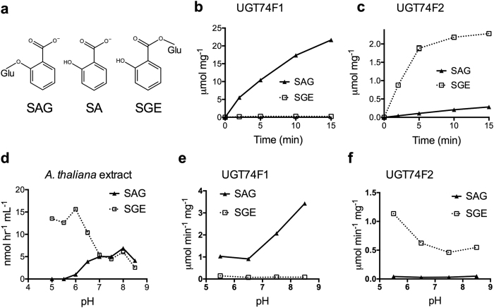
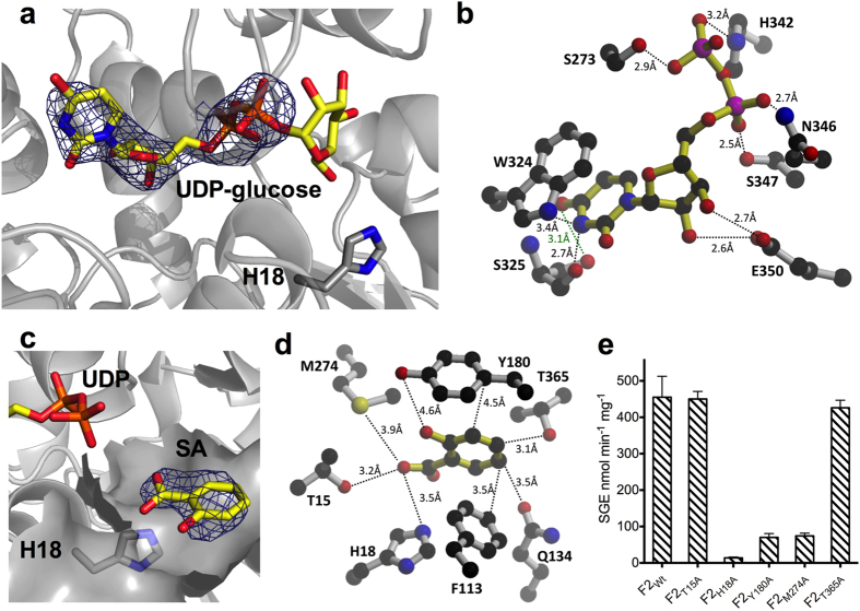
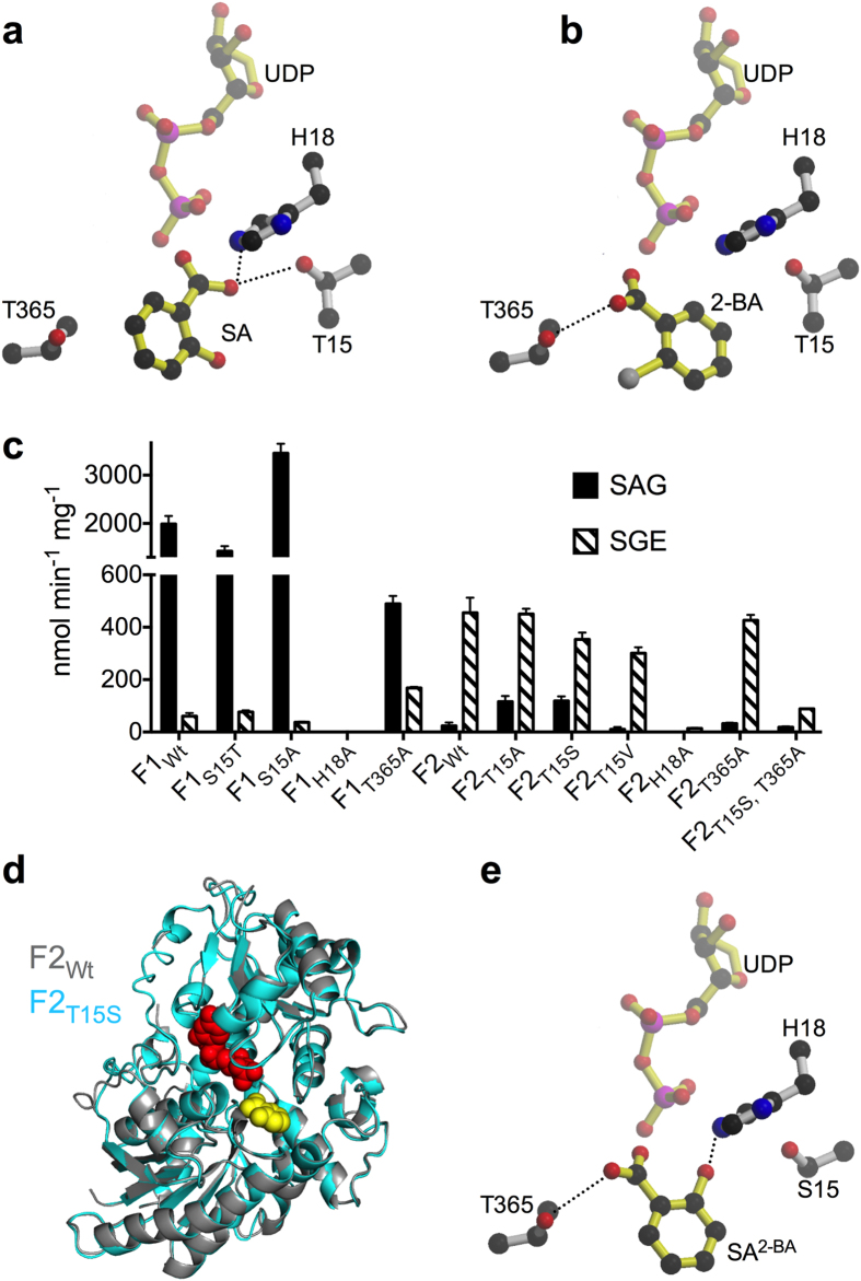
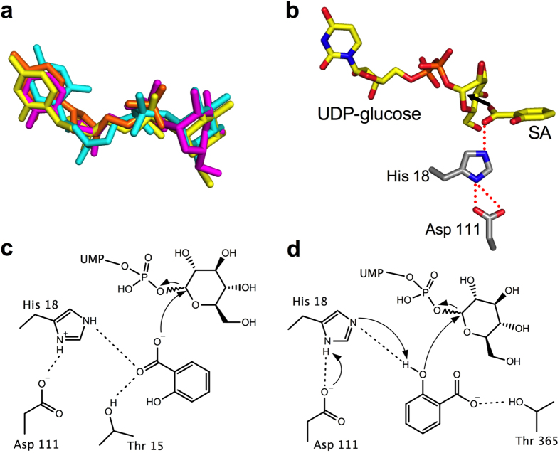
Salicylic acid (SA) is a signaling molecule utilized by plants in response to various stresses. Through conjugation with small organic molecules such as glucose, an inactive form of SA is generated which can be transported into and stored in plant vacuoles. In the model organism Arabidopsis thaliana, SA glucose conjugates are formed by two homologous enzymes (UGT74F1 and UGT74F2) that transfer glucose from UDP-glucose to SA. Despite being 77% identical and with conserved active site residues, these enzymes catalyze the formation of different products: UGT74F1 forms salicylic acid glucoside (SAG), while UGT74F2 forms primarily salicylic acid glucose ester (SGE). The position of the glucose on the aglycone determines how SA is stored, further metabolized, and contributes to a defense response. We determined the crystal structures of the UGT74F2 wild-type and T15S mutant enzymes, in different substrate/product complexes. On the basis of the crystal structures and the effect on enzyme activity of mutations in the SA binding site, we propose the catalytic mechanism of SGE and SAG formation and that SA binds to the active site in two conformations, with each enzyme selecting a certain binding mode of SA. Additionally, we show that two threonines are key determinants of product specificity.


Conflict of interest statement

The authors declare no competing financial interests.

Figures

Figure 1
Figure 1. Enzymatic formation of SAG and SGE.
(a) Structures of salicylic acid (SA), salicylic acid glucose ester (SGE), and salicylic acid glucoside (SAG); Glu represents the glucosyl moiety. Time-course product formation by the recombinant purified UGT74F1 (b) and UGT74F2 (c). The assays contained 1 μg protein, 5 mM UDP-glucose, 1 mM [7-14C]SA (1.5 μCi μmol−1), 50 mM Tris-HCl (pH 7.0), and 14 mM 2-mercaptoethanol in a total volume of 200 μL. (d) Formation of SAG and SGE in A. thaliana cell lysate over pH changes. The assay media included 50 μL cell lysate, 75 mM buffer, 10 mM UDP-glucose, and 0.14 mM [7-14C]SA (55 μCi μmol−1) in a total volume of 65 μL. pH-dependence of product formation by recombinant purified UGT74F1 (e) and UGT74F2 (f). Assays were performed as in (b) and (c) but in buffers of different pH (e and f). Except for (b) and (c) all assays are at 3 min time point. (bf) Each data point represents the average of 3 measurements.
Figure 2
Figure 2. Overview of UGT74F2 crystal structure.
(a) A chain of UGT74F2 in complex with UDP (red spheres) and SA (yellow spheres). N-terminal domain (residues 4 to 245) is colored teal with the C-terminal domain in grey. (b) Amino acid differences between UGT74F1 and UGT74F2 marked as blue on the UGT74F2 structure (see also Supplementary Fig. S3).
Figure 3
Figure 3. Ligand binding to UGT74F2.
(a) Omit electron density for UDP (1σ, blue mesh) shows clear density for UDP, but not for terminal glucose (UDP-glucose in yellow sticks). (b) Key residues interacting with UDP and potential hydrogen bonds. (c) Omit electron density (1σ, blue mesh) for SA. (d) Residues interacting with SA (for clarity we omitted residues Met 183, Val 184 and Trp 364). (e) SGE production by UGT74F2 mutants of the SA-binding site at 3 min in standard assay conditions. Error bars represent standard deviation from three measurements.
Figure 4
Figure 4. SA binding to UGT74F1 and UGT74F2.
(a) In wild-type UGT74F2 UDP/SA complex structure, the carboxyl group of SA interacts with Thr 15 and His 18, and faces UDP-glucose binding site. (b) In UGT74F2 wild-type and T15S UDP/2-BA complex structures 2-BA binds with the carboxyl group close to Thr 365. (c) SAG and SGE production of mutant proteins at 3 min in standard assay conditions compared to wild-type UGT74F1 and UGT74F2. Error bars represent standard deviation from three measurements. (d) Overlay of UGT74F2 wild-type (gray) and UGT74F2T15S (cyan) structures. (e) SA binding to the active site of UGT74F2T15S (or UGT74F1), modeled on the basis of 2-BA (SA2–BA, see b). The carboxyl group of modeled SA interacts with Thr 365, while SA hydroxyl interacts with His 18 and faces UDP. Dashed lines show hydrogen bond interactions.
Figure 5
Figure 5. Proposed catalytic mechanism for SA glucose conjugate formation.
(a) Modeled UDP-glucose molecules. Crystallographically observed UDP shown in orange, UDP-2-fluoro-glucose from PDB 2c1z shown in yellow, UDP-glucose modeled by Molecular Operating Environment is in cyan, and UDP-glucose modeled from incomplete electron density is shown in magenta. (b) View of proposed catalytic site for UGT74F2 showing modeled UDP-glucose, SA and the dyad His 18 - Asp 111. (c) View of catalytic site to form SGE, based on (b) and Fig. 4a: SA carboxyl oriented towards UDP-glucose by interactions with His 18 and Thr 15, performs an SN2 attack by the carboxylate oxygen to the anomeric carbon of UDP-glucose. (d) View of catalytic site for SAG formation, based on Fig. 4e: SA hydroxyl deprotonated by His 18 - Asp 111 catalytic dyad; SN2 attack by the adjacent aromatic hydroxyl to the anomeric carbon results in the formation of SAG, and the proton is released from His 18 to regenerate the active site.

References

    1. Raskin I., Skubatz H., Tang W. & Meeuse B. J. D. Salicylic Acid Levels in Thermogenic and Non-Thermogenic Plants. Ann. Bot. 66, 369–373 (1990).
    1. Malamy J., Carr J. P., Klessig D. F. & Raskin I. Salicylic Acid: a likely endogenous signal in the resistance response of tobacco to viral infection. Science 250, 1002–1004 (1990). - PubMed
    1. Delaney T. P. et al. A central role of salicylic Acid in plant disease resistance. Science 266, 1247–1250 (1994). - PubMed
    1. Métraux J. P. et al. Increase in salicylic Acid at the onset of systemic acquired resistance in cucumber. Science 250, 1004–1006 (1990). - PubMed
    1. Krinke O. et al. Phosphatidylinositol 4-kinase activation is an early response to salicylic acid in Arabidopsis suspension cells. Plant Physiol. 144, 1347–1359 (2007). - PMC - PubMed

Publication types

MeSH terms