

Osteoblast role in osteoarthritis pathogenesis
- PMID: 28425564
- PMCID: PMC5575507
- DOI: 10.1002/jcp.25969
Osteoblast role in osteoarthritis pathogenesis
Abstract
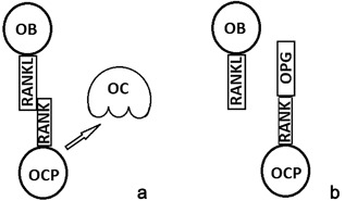
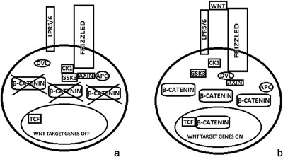
Even if osteoarthritis pathogenesis is still poorly understood, numerous evidences suggest that osteoblasts dysregulation plays a key role in osteoarthritis pathogenesis. An abnormal expression of OPG and RANKL has been described in osteoarthritis osteoblasts, which is responsible for abnormal bone remodeling and decreased mineralization. Alterations in genes expression are involved in dysregulation of osteoblast function, bone remodeling, and mineralization, leading to osteoarthritis development. Moreover, osteoblasts produce numerous transcription factors, growth factors, and other proteic molecules which are involved in osteoarthritis pathogenesis.
Keywords: bone; osteoarthritis; osteoblast.
© 2017 The Authors. Journal of Cellular Physiology Published by Wiley Periodicals, Inc.
Figures
Similar articles
-
RANKL/OPG ratio and DKK-1 expression in primary osteoblastic cultures from osteoarthritic and osteoporotic subjects.J Rheumatol. 2013 May;40(5):684-94. doi: 10.3899/jrheum.120845. Epub 2013 Mar 1. J Rheumatol. 2013. PMID: 23457386
-
Elevated levels of 15-lipoxygenase-1 contribute to the abnormal phenotypes of osteoblasts in human osteoarthritis.Life Sci. 2019 Dec 15;239:116980. doi: 10.1016/j.lfs.2019.116980. Epub 2019 Nov 5. Life Sci. 2019. PMID: 31704449
-
Strontium ranelate inhibits key factors affecting bone remodeling in human osteoarthritic subchondral bone osteoblasts.Bone. 2011 Sep;49(3):559-67. doi: 10.1016/j.bone.2011.06.005. Epub 2011 Jun 12. Bone. 2011. PMID: 21700005
-
Transcriptional regulation of bone and joint remodeling by NFAT.Immunol Rev. 2010 Jan;233(1):286-300. doi: 10.1111/j.0105-2896.2009.00849.x. Immunol Rev. 2010. PMID: 20193006 Free PMC article. Review.
-
Osteoblast Role in Rheumatic Diseases.Int J Mol Sci. 2017 Jun 15;18(6):1272. doi: 10.3390/ijms18061272. Int J Mol Sci. 2017. PMID: 28617323 Free PMC article. Review.
Cited by
-
Role of SIRT3 in bone homeostasis and its application in preventing and treating bone diseases.Front Pharmacol. 2023 Dec 20;14:1248507. doi: 10.3389/fphar.2023.1248507. eCollection 2023. Front Pharmacol. 2023. PMID: 38192409 Free PMC article. Review.
-
Long non-coding RNA H19 promotes osteogenic differentiation of human bone marrow-derived mesenchymal stem cells by regulating microRNA-140-5p/SATB2 axis.J Biosci. 2020;45:56. J Biosci. 2020. PMID: 32345782
-
Do Neuroendocrine Peptides and Their Receptors Qualify as Novel Therapeutic Targets in Osteoarthritis?Int J Mol Sci. 2018 Jan 26;19(2):367. doi: 10.3390/ijms19020367. Int J Mol Sci. 2018. PMID: 29373492 Free PMC article. Review.
-
The role of Clec11a in bone construction and remodeling.Front Endocrinol (Lausanne). 2024 Aug 12;15:1429567. doi: 10.3389/fendo.2024.1429567. eCollection 2024. Front Endocrinol (Lausanne). 2024. PMID: 39188913 Free PMC article. Review.
-
Tranexamic Acid Attenuates the Progression of Posttraumatic Osteoarthritis in Mice.Am J Sports Med. 2024 Mar;52(3):766-778. doi: 10.1177/03635465231220855. Epub 2024 Feb 2. Am J Sports Med. 2024. PMID: 38305280 Free PMC article.
References
-
- Abed, É , Chan, T. F. , Delalandre, A. , Martel‐Pelletier, J. , Pelletier, J. P. , & Lajeunesse, D. (2011). R‐spondins are newly recognized players in osteoarthritis that regulate Wnt signaling in osteoblasts. Arthritis & Rheumatism, 63, 3865–3875. - PubMed
-
- Abed, É , Couchourel, D. , Delalandre, A. , Duval, N. , Pelletier, J. P. , Martel‐Pelletier, J. , & Lajeunesse, D. (2014). Low sirtuin 1 levels in human osteoarthritis subchondral osteoblasts lead to abnormal sclerostin expression which decreases Wnt/β‐catenin activity. Bone, 59, 28–36. - PubMed
-
- Abed, É , Bouvard, B. , Martineau, X. , Jouzeau, J. Y. , Reboul, P. , & Lajeunesse, D. (2015). Elevated hepatocyte growth factor levels in osteoarthritis osteoblasts contribute to their altered response to bone morphogenetic protein‐2 and reduced mineralization capacity. Bone, 75, 111–119. - PubMed
Publication types
MeSH terms
LinkOut - more resources
Full Text Sources
Other Literature Sources
Medical
