. 2017 Jun;6(6):1557-1567.
doi: 10.1002/sctm.16-0398. Epub 2017 Apr 20.

Bone Marrow, Adipose, and Lung Tissue-Derived Murine Mesenchymal Stromal Cells Release Different Mediators and Differentially Affect Airway and Lung Parenchyma in Experimental Asthma

Affiliations

Bone Marrow, Adipose, and Lung Tissue-Derived Murine Mesenchymal Stromal Cells Release Different Mediators and Differentially Affect Airway and Lung Parenchyma in Experimental Asthma

Soraia C Abreu et al. Stem Cells Transl Med. 2017 Jun.

Abstract

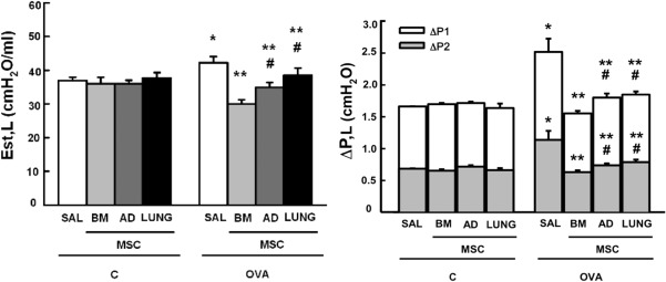
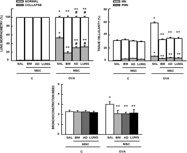
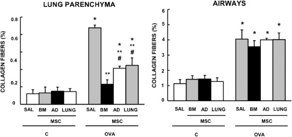
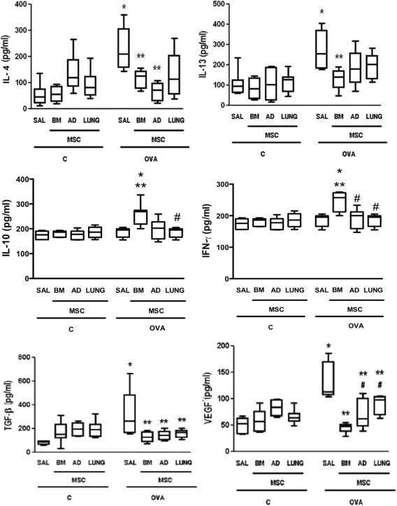
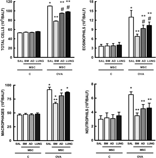
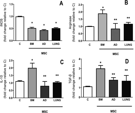
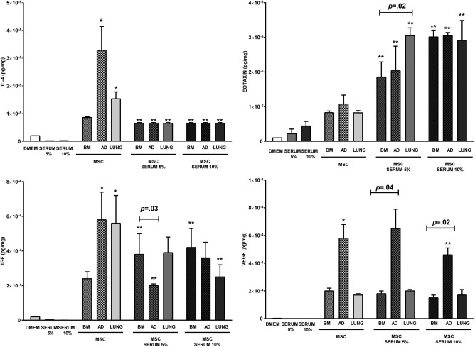
Mesenchymal stromal cells (MSCs) from different sources have differential effects on lung injury. To compare the effects of murine MSCs from bone marrow (BM), adipose tissue (AD), and lung tissue (LUNG) on inflammatory and remodeling processes in experimental allergic asthma, female C57BL/6 mice were sensitized and challenged with ovalbumin (OVA) or saline (C). Twenty-four hours after the last challenge, mice received either saline (50 µl, SAL), BM-MSCs, AD-MSCs, or LUNG-MSCs (105 cells per mouse in 50 µl total volume) intratracheally. At 1 week, BM-MSCs produced significantly greater reductions in resistive and viscoelastic pressures, bronchoconstriction index, collagen fiber content in lung parenchyma (but not airways), eosinophil infiltration, and levels of interleukin (IL)-4, IL-13, transforming growth factor (TGF)-β, and vascular endothelial growth factor (VEGF) in lung homogenates compared to AD-MSCs and LUNG-MSCs. Only BM-MSCs increased IL-10 and interferon (IFN)-γ in lung tissue. In parallel in vitro experiments, BM-MSCs increased M2 macrophage polarization, whereas AD-MSCs and LUNG-MSCs had higher baseline levels of IL-4, insulin-like growth factor (IGF), and VEGF secretion. Exposure of MSCs to serum specimens obtained from asthmatic mice promoted reductions in secretion of these mediators, particularly in BM-MSCs. Intratracheally administered BM-MSCs, AD-MSCs, and LUNG-MSCs were differentially effective at reducing airway inflammation and remodeling and improving lung function in the current model of allergic asthma. In conclusion, intratracheal administration of MSCs from BM, AD, and LUNG were differentially effective at reducing airway inflammation and remodeling and improving lung function comparably reduced inflammation and fibrogenesis in this asthma model. However, altered lung mechanics and lung remodeling responded better to BM-MSCs than to AD-MSCs or LUNG-MSCs. Moreover, each type of MSC was differentially affected in a surrogate in vitro model of the in vivo lung environment. Stem Cells Translational Medicine 2017;6:1557-1567.

Keywords: Asthma; Fibrosis; Inflammation; Macrophage; Mesenchymal stromal cells.


Figures

Figure 1
Figure 1
Lung mechanics. Static lung elastance (Est, L) and resistive (ΔP1, gray bar), viscoelastic (ΔP2, white bar), and total pressures (ΔPtot = ΔP1 + ΔP2). C: mice sensitized and challenged with saline; OVA: mice sensitized and challenged with ovalbumin; SAL: mice sensitized and challenged with ovalbumin and treated with saline; BM‐, AD‐, and LUNG‐MSCs: mice treated with MSCs derived from bone marrow, adipose, and lung tissues, respectively, 24 hours after the last challenge (105 cells per mice). *Significantly different from C‐SAL (p < .05). **Significantly different from OVA‐SAL (p < .05). #Significantly different from OVA‐BM‐MSCs (p < .05). Abbreviations: AD, adipose tissue; BM, bone marrow; LUNG, lung tissue; MSCs, mesenchymal stromal cells; OVA, ovalbumin; SAL, saline.
Figure 2
Figure 2
Morphometric parameters. Lung morphometry: fractional area of normal (white bar) and collapsed alveoli (gray bar); Tissue cellularity: fractional area of mononuclear (MN, white bar) and polymorphonuclear (PMN, gray bar) cells; and Bronchoconstriction index. Bars are means + SD of six animals per group. C: mice sensitized and challenged with saline; OVA: mice sensitized and challenged with ovalbumin; SAL: mice sensitized and challenged with ovalbumin and treated with saline; BM‐, AD‐, and LUNG‐MSCs: mice treated with MSCs derived from bone marrow, adipose, and lung tissues, respectively, 24 hours after the last challenge (105 cells per mice). *Significantly different from C‐SAL (p < .05). **Significantly different from OVA‐SAL (p < .05). #Significantly different from OVA‐BM‐MSCs (p < .05). Abbreviations: AD, adipose tissue; BM, bone marrow; LUNG, lung tissue; MSCs, mesenchymal stromal cells; OVA, ovalbumin; SAL, saline.
Figure 3
Figure 3
Collagen fiber content in lung parenchyma and airways. Bars are means + SD of six animals per group. C: mice sensitized and challenged with saline; OVA: mice sensitized and challenged with ovalbumin; SAL: mice sensitized and challenged with ovalbumin and treated with saline; BM‐, AD‐, and LUNG‐MSCs: mice treated with MSCs derived from bone marrow, adipose, and lung tissues, respectively, 24 hours after the last challenge (105 cells per mice). *Significantly different from C‐SAL (p < .05). **Significantly different from OVA‐SAL (p < .05). #Significantly different from OVA‐BM‐MSCs (p < .05). Abbreviations: AD, adipose tissue; BM, bone marrow; LUNG, lung tissue; MSCs, mesenchymal stromal cells; OVA, ovalbumin; SAL, saline.
Figure 4
Figure 4
Levels of interleukin (IL)‐4, IL‐13, IL‐10, interferon (IFN)‐γ, transforming growth factor (TGF)‐β, and vascular endothelial growth factor in lung tissue. Boxes show the interquartile (25%–75%) range, whiskers encompass the range (minimum–maximum), and horizontal lines represent the median in six animals per group. C: mice sensitized and challenged with saline; OVA: mice sensitized and challenged with ovalbumin; SAL: mice sensitized and challenged with ovalbumin and treated with saline; BM, AD, and LUNG‐MSCs: mice treated with MSCs derived from bone marrow, adipose, and lung tissues, respectively, 24 hours after the last challenge (105 cells per mice). *Significantly different from C‐SAL (p < .05). **Significantly different from OVA‐SAL (p < .05). #Significantly different from OVA‐BM‐MSCs (p < .05). Abbreviations: AD, adipose tissue; BM, bone marrow; IL, interleukin; iNOS, inducible nitric oxide synthase; LUNG, lung tissue; MSCs, mesenchymal stromal cells; OVA, ovalbumin; SAL, saline; TGF, transforming growth factor.
Figure 5
Figure 5
Number of total cells, eosinophils, macrophages, and neutrophils in bronchoalveolar lavage fluid. Bars are means + SD of six animals per group. C: mice sensitized and challenged with saline; OVA: mice sensitized and challenged with ovalbumin; SAL: mice sensitized and challenged with ovalbumin and treated with saline; BM, AD, and LUNG‐MSCs: mice treated with MSCs derived from bone marrow, adipose, and lung tissues, respectively, 24 hours after the last challenge (105 cells per mice). *Significantly different from C‐SAL (p < .05). **Significantly different from OVA‐SAL (p < .05). #Significantly different from OVA‐BM‐MSCs (p < .05). Abbreviations: AD, adipose tissue; BM, bone marrow; LUNG, lung tissue; MSCs, mesenchymal stromal cells; OVA, ovalbumin; SAL, saline.
Figure 6
Figure 6
In vitro assay. RAW 264.7, a macrophage cell line, was cocultured with different MSCs for 24 hours. Relative gene expression of iNOS (A), arginase (B), IL‐10 (C), and (D) TGF‐β was calculated as a ratio of the average gene expression levels compared with the reference gene (36B4) and expressed as fold changes relative to C group (noncontacting cell cocultures, 105 RAW 264.7/well). Bars are means + SD of five wells per condition. BM‐, AD‐, and LUNG‐MSCs: macrophages were co‐cultured in transwell plates with MSCs derived from bone marrow, adipose, and lung tissues (105 MSCs per well). *Significantly different from C (p < .05). **Significantly different from BM‐MSCs (p < .05). Abbreviations: AD, adipose tissue; BM, bone marrow; IL, interleukin; iNOS, inducible nitric oxide synthase; LUNG, lung tissue; MSCs, mesenchymal stromal cells; OVA, ovalbumin; SAL, saline; TGF, transforming growth factor.
Figure 7
Figure 7
Levels of interleukin (IL)‐4, eotaxin, IGF, and vascular endothelial growth factor in the coculture medium after BM‐, AD‐, and LUNG‐MSC stimulation with serum obtained from OVA mice. DMEM: Dulbecco's Modified Eagle's Medium; SERUM 5%: serum obtained from OVA mice at a concentration of 5%; SERUM 10%: serum obtained from OVA mice at a concentration of 10%; BM‐, AD‐, and LUNG‐: MSCs from bone marrow, adipose, and lung tissues (105 cells per well), respectively, unstimulated or stimulated with 5% or 10% serum obtained from OVA mice during 24 hours. *Significantly different from BM‐MSCs (p < .05). **Significantly different from unstimulated MSCs (p < .05). Abbreviations: IGF, insulin‐like growth factor; MSCs, mesenchymal stromal cells.

Similar articles

Cited by

References

    1. Al‐Muhsen S, Johnson JR, Hamid Q. Remodeling in asthma. J Allergy Clin Immunol 2011;128:451–462; quiz 463–454. - PubMed
    1. Cruz FF, Borg ZD, Goodwin M et al. Systemic administration of human bone marrow‐derived mesenchymal stromal cell extracellular vesicles ameliorates aspergillus hyphal extract‐induced allergic airway inflammation in immunocompetent mice. Stem Cell Transl Med 2015;4:1302–1316. - PMC - PubMed
    1. Fu QL, Chow YY, Sun SJ et al. Mesenchymal stem cells derived from human induced pluripotent stem cells modulate T‐cell phenotypes in allergic rhinitis. Allergy 2012;67:1215–1222. - PMC - PubMed
    1. Lathrop MJ, Brooks EM, Bonenfant NR et al. Mesenchymal stromal cells mediate Aspergillus hyphal extract‐induced allergic airway inflammation by inhibition of the th17 signaling pathway. Stem Cells Transl Med 2014;3:194–205. - PMC - PubMed
    1. Jang HJ, Cho KS, Park HY et al. Adipose tissue‐derived stem cells for cell therapy of airway allergic diseases in mouse. Acta Histochem 2011;113:501–507. - PubMed

Publication types

Substances