

Nck2, an unexpected regulator of adipogenesis
- PMID: 28425845
- PMCID: PMC5477721
- DOI: 10.1080/21623945.2017.1291102
Nck2, an unexpected regulator of adipogenesis
Abstract
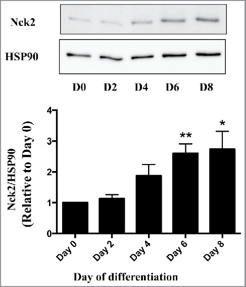
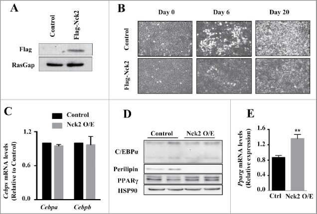
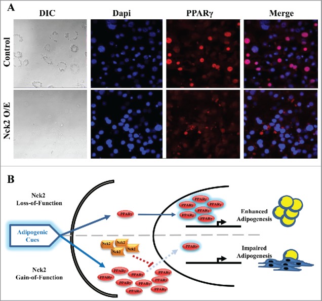
The regulation of adipose tissue expansion by adipocyte hypertrophy and/or hyperplasia is the topic of extensive investigations given the potential differential contribution of the 2 processes to the development of numerous chronic diseases associated with obesity. We recently discovered that the loss-of-function of the Src homology domain-containing protein Nck2 in mice promotes adiposity accompanied with adipocyte hypertrophy and impaired function, and enhanced adipocyte differentiation in vitro. Moreover, in severely-obese human's adipose tissue, we found that Nck2 expression is markedly downregulated. In this commentary, our goal is to expand upon additional findings providing further evidence for a unique Nck2-dependent mechanism regulating adipogenesis. We propose that Nck2 should be further investigated as a regulator of the reliance of white adipose tissue on hyperplasia versus hypertrophy during adipose tissue expansion, and hence, as a potential novel molecular target in obesity.
Keywords: 3T3-L1 and SGBS preadipocytes; Adipogenesis; PERK activation and signaling; PPARγ regulation; Src homology adaptor protein Nck2; adipocyte differentiation.
Figures
Similar articles
-
Nck2 Deficiency in Mice Results in Increased Adiposity Associated With Adipocyte Hypertrophy and Enhanced Adipogenesis.Diabetes. 2016 Sep;65(9):2652-66. doi: 10.2337/db15-1559. Epub 2016 Jun 20. Diabetes. 2016. PMID: 27325288
-
The role and possible mechanism of lncRNA U90926 in modulating 3T3-L1 preadipocyte differentiation.Int J Obes (Lond). 2017 Feb;41(2):299-308. doi: 10.1038/ijo.2016.189. Epub 2016 Oct 26. Int J Obes (Lond). 2017. PMID: 27780975 Free PMC article.
-
The anti-angiogenic herbal extract from Melissa officinalis inhibits adipogenesis in 3T3-L1 adipocytes and suppresses adipocyte hypertrophy in high fat diet-induced obese C57BL/6J mice.J Ethnopharmacol. 2016 Feb 3;178:238-50. doi: 10.1016/j.jep.2015.12.015. Epub 2015 Dec 15. J Ethnopharmacol. 2016. PMID: 26702505
-
Harnessing adipogenesis to prevent obesity.Adipocyte. 2019 Dec;8(1):98-104. doi: 10.1080/21623945.2019.1583037. Epub 2019 Mar 8. Adipocyte. 2019. PMID: 30848691 Free PMC article. Review.
-
Fifty shades of white: Understanding heterogeneity in white adipose stem cells.Adipocyte. 2017 Jul 3;6(3):205-216. doi: 10.1080/21623945.2017.1372871. Epub 2017 Sep 12. Adipocyte. 2017. PMID: 28949833 Free PMC article. Review.
Cited by
-
Nck1 Deficiency Impairs Adipogenesis by Activation of PDGFRα in Preadipocytes.iScience. 2018 Aug 31;6:22-37. doi: 10.1016/j.isci.2018.07.010. Epub 2018 Jul 19. iScience. 2018. PMID: 30240612 Free PMC article.
-
Ca2+ entry via TRPC1 is essential for cellular differentiation and modulates secretion via the SNARE complex.J Cell Sci. 2019 Jul 1;132(13):jcs231878. doi: 10.1242/jcs.231878. J Cell Sci. 2019. PMID: 31182642 Free PMC article.
-
20 Years with SGBS cells - a versatile in vitro model of human adipocyte biology.Int J Obes (Lond). 2022 Nov;46(11):1939-1947. doi: 10.1038/s41366-022-01199-9. Epub 2022 Aug 19. Int J Obes (Lond). 2022. PMID: 35986215 Free PMC article. Review.
-
Father's adolescent body silhouette is associated with offspring asthma, lung function and BMI through DNA methylation.Commun Biol. 2025 May 24;8(1):796. doi: 10.1038/s42003-025-08121-9. Commun Biol. 2025. PMID: 40410506 Free PMC article.
References
-
- Brook CG, Lloyd JK, Wolf OH. Relation between age of onset of obesity and size and number of adipose cells. Br Med J 1972; 2(5804):25-7; PMID:5015967; https://doi.org/10.1136/bmj.2.5804.25 - DOI - PMC - PubMed
-
- Wueest S, Rapold RA, Rytka JM, Schoenle EJ, Konrad D. Basal lipolysis, not the degree of insulin resistance, differentiates large from small isolated adipocytes in high-fat fed mice. Diabetologia 2009; 52(3):541-6; PMID:19048227; https://doi.org/10.1007/s00125-008-1223-5 - DOI - PubMed
-
- Skurk T, Alberti-Huber C, Herder C, Hauner H. Relationship between adipocyte size and adipokine expression and secretion. J Clin Endocrinol Metab 2007; 92(3):1023-33; PMID:17164304; https://doi.org/10.1210/jc.2006-1055 - DOI - PubMed
-
- Cotillard A, Poitou C, Torcivia A, Bouillot JL, Dietrich A, Kloting N, Grégoire C, Lolmede K, Blüher M, Clément K. Adipocyte size threshold matters: link with risk of type 2 diabetes and improved insulin resistance after gastric bypass. J Clin Endocrinol Metab 2014; 99(8):E1466-70; PMID:24780048; https://doi.org/10.1210/jc.2014-1074 - DOI - PubMed
-
- Rosen ED, MacDougald OA. Adipocyte differentiation from the inside out. Nat Rev Mol Cell Biol 2006; 7(12):885-96; PMID:17139329; https://doi.org/10.1038/nrm2066 - DOI - PubMed
Publication types
MeSH terms
Substances
Grants and funding
LinkOut - more resources
Full Text Sources
Other Literature Sources
Miscellaneous