Review
. 2017 Apr 20;18(4):869.
doi: 10.3390/ijms18040869.

Hepatitis E Virus Genotypes and Evolution: Emergence of Camel Hepatitis E Variants

Affiliations
Review

Hepatitis E Virus Genotypes and Evolution: Emergence of Camel Hepatitis E Variants

Siddharth Sridhar et al. Int J Mol Sci. .

Abstract

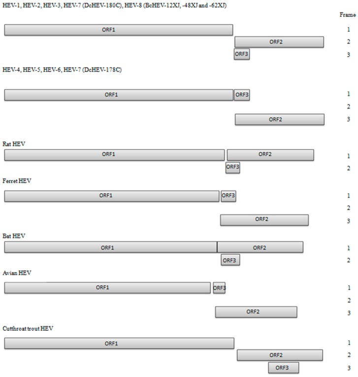
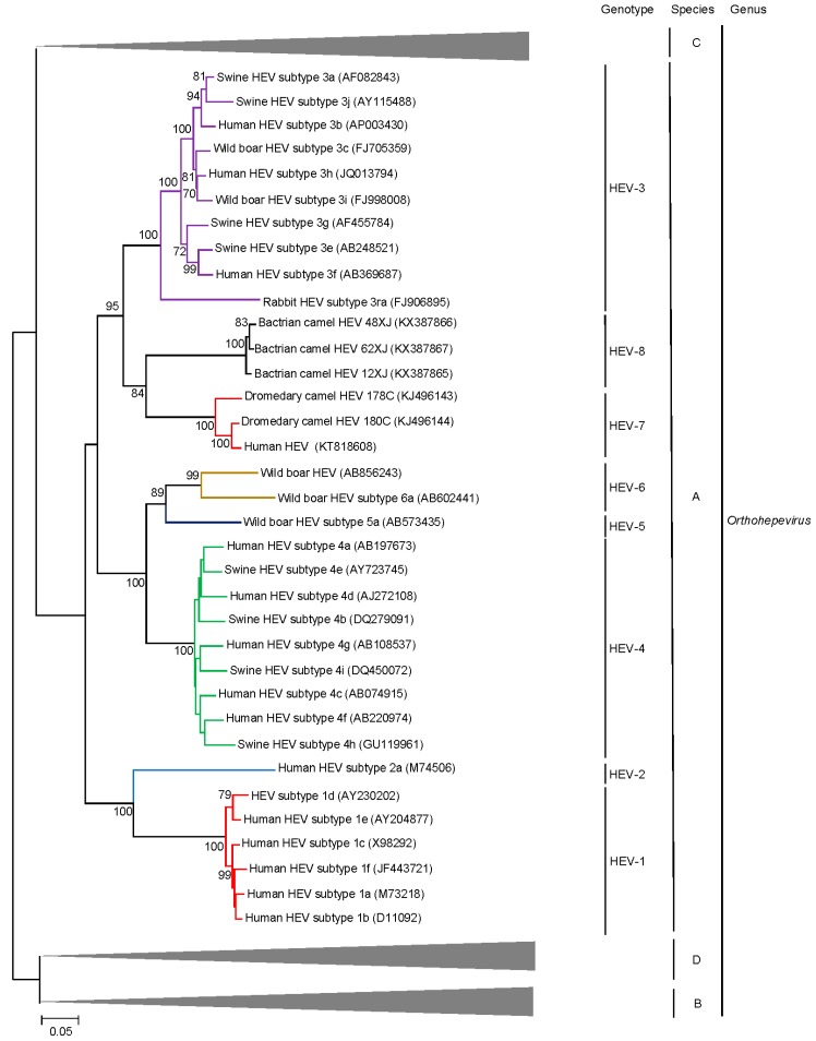
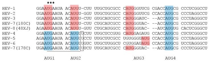
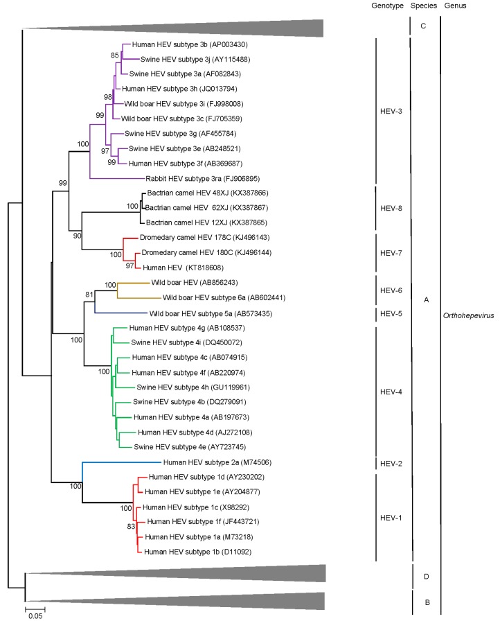
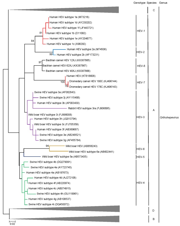
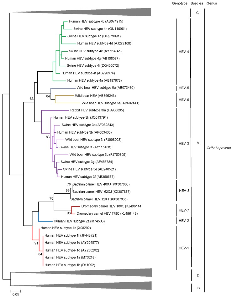
Hepatitis E virus (HEV) is a major cause of viral hepatitis globally. Zoonotic HEV is an important cause of chronic hepatitis in immunocompromised patients. The rapid identification of novel HEV variants and accumulating sequence information has prompted significant changes in taxonomy of the family Hepeviridae. This family includes two genera: Orthohepevirus, which infects terrestrial vertebrates, and Piscihepevirus, which infects fish. Within Orthohepevirus, there are four species, A-D, with widely differing host range. Orthohepevirus A contains the HEV variants infecting humans and its significance continues to expand with new clinical information. We now recognize eight genotypes within Orthohepevirus A: HEV1 and HEV2, restricted to humans; HEV3, which circulates among humans, swine, rabbits, deer and mongooses; HEV4, which circulates between humans and swine; HEV5 and HEV6, which are found in wild boars; and HEV7 and HEV8, which were recently identified in dromedary and Bactrian camels, respectively. HEV7 is an example of a novel genotype that was found to have significance to human health shortly after discovery. In this review, we summarize recent developments in HEV molecular taxonomy, epidemiology and evolution and describe the discovery of novel camel HEV genotypes as an illustrative example of the changes in this field.

Keywords: Bactrian camel; dromedary camel; genotypes; hepatitis; hepatitis E; molecular epidemiology; molecular evolution; swine; taxonomy; zoonosis.


Conflict of interest statement

The authors declare no conflicts of interest.

Figures

Figure 1
Figure 1
Predicted genomic organization of HEV-1 to HEV-8, considering the reading frame of ORF1 as frame 1. Reading frames of ORF2 and ORF3 are labeled relative to ORF1 reading frame. HEV-1 (M73218); HEV-2 (M74506); HEV-3 (AP003430); HEV-4 (AJ272108); HEV-5 (AB573435); HEV-6 (AB602441); HEV-7 strain DcHEV-178C (KJ496143); HEV-7 strain DcHEV-180C (KJ496144), HEV-8 (KX387865).
Figure 2
Figure 2
Phylogenetic analyses of ORF1/ORF2 proteins excluding the hypervariable region (HVR) of HEVs within the genus Orthohepevirus (Species A to D). The tree was constructed using maximum likelihood method and the optimal substitution model of JTT + G + I was used. In total, 2282 acid positions (amino acid residues 1–706 and 789–2409, numbered with reference to GenBank sequence M73218) were included in the analyses. The scale bar indicates the estimated number of substitutions per 20 amino acids. GenBank accession numbers are shown in brackets.
Figure 3
Figure 3
Alignment of nucleotide sequences showing potential start codons for ORF2 and ORF3 in HEV-1 to HEV-8. Potential start codons of ORF3 are shaded by red boxes, and those of ORF2 by blue boxes. The inserted U residues are indicated by open box. The stop codon of ORF1 is marked with asterisks. The conserved cis-reactive element with the sequence UGAAUAACAUGU is underlined, which might serve as promoter for the synthesis of the subgenomic mRNA for the ORF2 and ORF3.
Figure 4
Figure 4
Phylogenetic analyses of ORF1of HEVs within the genus Orthohepevirus (Species A to D). The tree was constructed using maximum likelihood method and the optimal substitution model of JTT + G + I + F was used. Amino acid residues 1–1743 in ORF1, numbered with reference to GenBank sequence M73218, were included in the analyses. The scale bar indicates the estimated number of substitutions per 20 amino acids. GenBank accession numbers are shown in brackets.
Figure 5
Figure 5
Phylogenetic analyses of ORF2 of HEVs within the genus Orthohepevirus (Species A to D). The tree was constructed using maximum likelihood method and the optimal substitution model of JTT + G + I was used. Amino acid residues 1–660 in ORF2, numbered with reference to GenBank sequence M73218, were included in the analyses. The scale bar indicates the estimated number of substitutions per 50 amino acids. GenBank accession numbers are shown in brackets.
Figure 6
Figure 6
Phylogenetic analyses of ORF3 of HEVs within the genus Orthohepevirus (Species A to D). The tree was constructed using maximum likelihood method and the optimal substitution model of JTT + G was used. Amino acid residues 10–123 in ORF3, numbered with reference to GenBank sequence M73218, were included in the analyses. The scale bar indicates the estimated number of substitutions per 20 amino acids. GenBank accession numbers are shown in brackets.

References

    1. WHO Hepatitis E Fact Sheet. [(accessed on 15 February 2017)]; Available online: http://www.who.int/mediacentre/factsheets/fs280/en/
    1. Ren X., Wu P., Wang L., Geng M., Zeng L., Zhang J., Xia N., Lai S., Dalton H.R., Cowling B.J., et al. Changing epidemiology of hepatitis A and hepatitis E viruses in China, 1990–2014. Emerg. Infect. Dis. 2017;23:276–279. doi: 10.3201/eid2302.161095. - DOI - PMC - PubMed
    1. Jin H., Zhao Y., Zhang X., Wang B., Liu P. Case-fatality risk of pregnant women with acute viral hepatitis type E: A systematic review and meta-analysis. Epidemiol. Infect. 2016;144:2098–2106. doi: 10.1017/S0950268816000418. - DOI - PMC - PubMed
    1. Kamar N., Selves J., Mansuy J.M., Ouezzani L., Peron J.M., Guitard J., Cointault O., Esposito L., Abravanel F., Danjoux M., et al. Hepatitis E virus and chronic hepatitis in organ-transplant recipients. N. Engl. J. Med. 2008;358:811–817. doi: 10.1056/NEJMoa0706992. - DOI - PubMed
    1. Giordani M.T., Fabris P., Brunetti E., Goblirsch S., Romano L. Hepatitis E and lymphocytic leukemia in Man, Italy. Emerg. Infect. Dis. 2013;19:2054–2056. doi: 10.3201/eid1912.130521. - DOI - PMC - PubMed