. 2017 Apr 20;8(4):e2753.
doi: 10.1038/cddis.2017.171.

Biological activity of tumor-treating fields in preclinical glioma models

Affiliations

Biological activity of tumor-treating fields in preclinical glioma models

Manuela Silginer et al. Cell Death Dis. .

Abstract

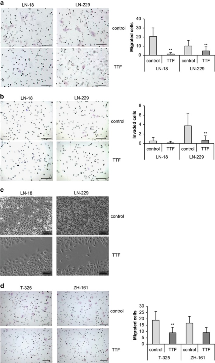
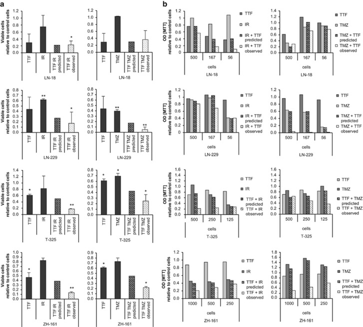
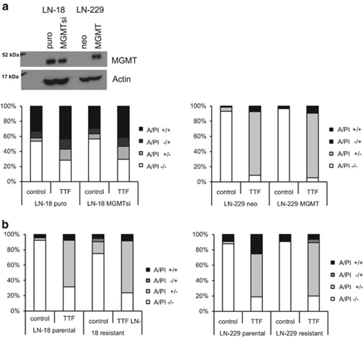
Glioblastoma is the most common and aggressive form of intrinsic brain tumor with a very poor prognosis. Thus, novel therapeutic approaches are urgently needed. Tumor-treating fields (TTFields) may represent such a novel treatment option. The aim of this study was to investigate the effects of TTFields on glioma cells, as well as the functional characterization of the underlying mechanisms. Here, we assessed the anti-glioma activity of TTFields in several preclinical models. Applying TTFields resulted in the induction of cell death in a frequency- and intensity-dependent manner in long-term glioma cell lines, as well as glioma-initiating cells. Cell death occurred in the absence of caspase activation, but involved autophagy and necroptosis. Severe alterations in cell cycle progression and aberrant mitotic features, such as poly- and micronucleation, preceded the induction of cell death. Furthermore, exposure to TTFields led to reduced migration and invasion, which are both biological hallmarks of glioma cells. The combination of TTFields with irradiation or the alkylating agent, temozolomide (TMZ), resulted in additive or synergistic effects, and the O6-methyl-guanine DNA methyltransferase status did not influence the efficacy of TTFields. Importantly, TMZ-resistant glioma cells were responsive to TTFields application, highlighting the clinical potential of this therapeutic approach. In summary, our results indicate that TTFields induce autophagy, as well as necroptosis and hamper the migration and invasiveness of glioma cells. These findings may allow for a more detailed clinical evaluation of TTFields beyond the clinical data available so far.


Conflict of interest statement

MW has received research grants from Actelion, Alpinia Institute, Bayer, Isarna, MSD, Merck Serono, Piqur and Roche and honoraria for lectures or advisory board participation from Celldex, Isarna, Magforce, MSD, Merck Serono, Pfizer, Roche and Teva. RS is the principal investigator for the pivotal clinical trials on TTFields in newly diagnosed and recurrent glioblastoma. RS served as an advisor to Novocure (non-remunerated), and has received travel assistance for trial associated activities, data review and data presentation at scientific meetings. PR has received honoraria for advisory boards and lectures from BMS, Roche, MSD, Novartis and Molecular Partners. MS declares no conflict of interest.

Figures

Figure 1
Figure 1
TTFields induce cell death in an intensity- and frequency-dependent manner in human glioma cells. (a) LN-18, LN-229, T-325 or ZH-161 cells were left untreated or exposed to TTFields (TTF; 2 V/cm) at the indicated frequencies for 48 h. Viable cell counts were obtained using trypan blue exclusion (*P<0.05). (b) The human LTC LN-18 or LN-229 (top) or T-325 or ZH-161 GIC (bottom) were exposed to increasing intensities of TTFields as indicated for 48 h (LTC) or 72 h (GIC). Cell death was assessed by annexin V/PI staining. (c) The LTC LN-428, LN-319, A172, U87MG, T98G or LN-308 (top) and the GIC T-269, S-24 or ZH-305 (bottom) were exposed to TTFields (2 V/cm) or not for 48 or 72 h, respectively, and subsequently analyzed for cell death by annexin V/PI staining
Figure 2
Figure 2
TTFields induce cell death via autophagy. (a) LN-18, LN-229 or ZH-161 cells were left untreated or exposed to TTFields (TTF, 3 V/cm), staurosporine (Stauro, 1 μM), zVAD-fmk (10 μM) or combinations thereof for 6 h. DEVD-amc cleaving activity was determined fluorometrically (**P<0.01). (b) Whole-cell lysates of LN-18, LN-229 or ZH-161 cells, untreated or exposed to TTF (3 V/cm) or staurosporine (1 μM) for 48 h, were analyzed for full-length and cleaved caspase-3 and LC3A/B protein levels. Actin was used as a loading control. (c) LN-18 or ZH-161 cells were left untreated or exposed to TTFields (TTF, 3 V/cm) or Stauro (1 μM) for 48 h and then monitored by electron microscopy. Autophagosomes are indicated with white arrows, white arrowheads point to mitochondria with swollen matrices, the white star to the dilated endoplasmatic reticulum, black arrows to membrane blebs and black arrowheads to condensed DNA along the nucleus (scale bar, 2 μm). (d) LN-18 or ZH-161 cells were treated with 3-methyladenine (3-MA, 1 mM) for 60 min followed by TTFields (2 V/cm) or not for 72 h. Cell death was assessed by annexin V/PI staining. (e) LN-18 or ZH-161 cells were treated with necrostatin-1 (Nec-1; 100 μM) for 60 min followed by TTFields (3 V/cm) or not for 72 h. Cell death was assessed by annexin V/PI staining
Figure 3
Figure 3
TTFields interfere with cell cycle progression. (a) LN-18 or LN-229 cells were exposed to TTFields (2 V/cm) for 24 or 48 h as indicated. Cell cycle analysis was performed by flow cytometry and cell cycle distribution is shown in bar graphs, as well as flow cytometry profiles. (b) LN-18 cells were left untreated or exposed to TTFields (2 V/cm) for 24 h. Actin (phalloidin staining, green), beta III tubulin (red) and nuclei (DAPI, blue) were analyzed by immunofluorescence. Different magnifications are shown. An isotype control for anti-beta III tubulin is included at the bottom. Accumulations of nuclear actin are indicated with arrows, arrowheads point to altered cell shape and stars to nuclear aberrations (scale bar, 10 μm)
Figure 4
Figure 4
TTFields reduce migration and invasion of glioma cells. (a and b) LN-18 or LN-229 cells were left untreated or exposed to TTFields (2 V/cm) for 24 h. Subsequently, modified migration/matrigel invasion Boyden chamber assays were performed to analyze migration (a) or invasion (b). Data are expressed as mean cells per field of vision (FoV) and exemplary photographs of migrated or invaded LN-18 or LN-229 cells are shown (scale bar, 100 μm). (c) A scratch wound-healing assay was performed during which LN-18 or LN-229 cells were left untreated or exposed to TTFields (2 V/cm). After 24 h, the gap distance was imaged on a microscope and representative images are shown (scale bar, 100 μm). (d) T-325 or ZH-161 cells were left untreated or exposed to TTFields (2 V/cm) for 24 h. Subsequently, a modified migration Boyden chamber assay was performed. Data are expressed as mean cells per FoV and exemplary photographs of migrated cells preexposed to TTFields or not are shown (scale bar, 100 μm). (**P<0.01)
Figure 5
Figure 5
TTFields act synergistically with irradiation or TMZ to reduce acute and clonogenic survival. (a) LN-18, LN-229, T-325 or ZH-161 cells were left untreated or irradiated (LN-18, LN-229, 3 Gy; T-325 and ZH-161, 5 Gy) followed or not by TTFields (2 V/cm) for 24 h (left panel). Alternatively, the cells were exposed to DMSO control or TMZ (LN-18, 100 μM; LN-229, 5 μM; T-325, 200 μM; ZH-161, 25 μM) paralleled by TTFields exposure (2 V/cm) for 24 h or not (right panel). After another 48 h, viable cells were counted by Trypan blue exclusion test (*P<0.05; **P<0.01, relative to control cells). (b) Cells, treated as in a, were seeded at the indicated densities and clonogenic survival was assessed by 3-(4,5-dimethylthiazol-2-yl)-2,5-diphenyltetrazolium bromide (MTT) assay after 14 days
Figure 6
Figure 6
TTFields induce cell death in a MGMT-independent manner. (a) Whole-cell lysates of LN-18 puro or MGMTsi, or LN-229 neo or MGMT cells were analyzed for MGMT expression by immunoblot. Actin was used as a control (top). The cells were exposed to TTFields (2 V/cm) or not for 48 h and analyzed for cell death by annexin V/PI staining. (b) LN-18 parental or TMZ-resistant, or LN-229 parental or TMZ-resistant cells were exposed to TTFields (2 V/cm) or not. After 48 h cell death was analyzed by annexin V/PI staining

References

    1. Weller M, van den Bent M, Hopkins K, Tonn JC, Stupp R, Falini A et al. EANO guideline for the diagnosis and treatment of anaplastic gliomas and glioblastoma. Lancet Oncol 2014; 15: e395–e403. - PubMed
    1. Pless M, Weinberg U. Tumor treating fields: concept, evidence and future. Expert Opin Investig Drugs 2011; 20: 1099–1106. - PubMed
    1. Davies AM, Weinberg U, Palti Y. Tumor treating fields: a new frontier in cancer therapy. Ann N Y Acad Sci 2013; 1291: 86–95. - PubMed
    1. Kirson ED, Gurvich Z, Schneiderman R, Dekel E, Itzhaki A, Wasserman Y et al. Disruption of cancer cell replication by alternating electric fields. Cancer Res 2004; 64: 3288–3295. - PubMed
    1. Kirson ED, Dbaly V, Tovarys F, Vymazal J, Soustiel JF, Itzhaki A et al. Alternating electric fields arrest cell proliferation in animal tumor models and human brain tumors. Proc Natl Acad Sci USA 2007; 104: 10152–10157. - PMC - PubMed

MeSH terms