

Hippocampal electrical stimulation disrupts associative learning when targeted at dentate spikes
- PMID: 28426128
- PMCID: PMC5509848
- DOI: 10.1113/JP274023
Hippocampal electrical stimulation disrupts associative learning when targeted at dentate spikes
Abstract
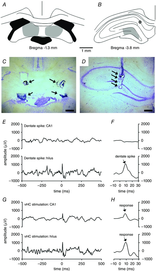
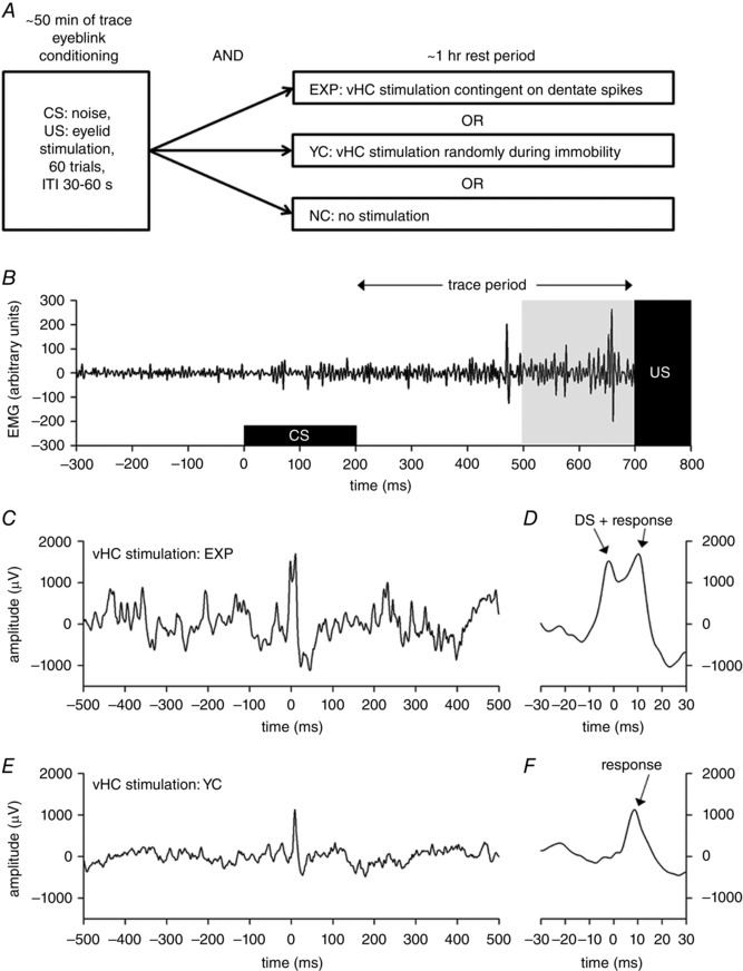
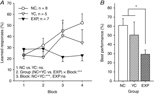
Key points: Dentate spikes are fast fluctuations of hilar local-field potentials that take place during rest and are thought to reflect input arriving from the entorhinal cortex to the hippocampus. During dentate spikes, neuronal firing in hippocampal input (dentate gyrus) and output (CA1/CA3) regions is uncoupled. To date, the behavioural significance of dentate spikes is unknown. Here, we provide evidence that disrupting the dentate spike-related uncoupling of the dentate gyrus and the CA1/CA3 subregions for 1 h after training retards associative learning. We suggest dentate spikes play a significant role in memory consolidation.
Abstract: Hippocampal electrophysiological oscillations, namely theta and ripples, have been implicated in encoding and consolidation of new memories, respectively. According to existing literature, hippocampal dentate spikes are prominent, short-duration (<30 ms), large-amplitude (∼2-4 mV) fluctuations in hilar local-field potentials that take place during awake immobility and sleep. Interestingly, previous studies indicate that during dentate spikes dentate gyrus granule cells increase their firing while firing of CA1 pyramidal cells are suppressed, thus resulting in momentary uncoupling of the two hippocampal subregions. To date, the behavioural significance of dentate spikes is unknown. Here, to study the possible role of dentate spikes in learning, we trained adult male Sprague-Dawley rats in trace eyeblink classical conditioning. For 1 h immediately following each conditioning session, one group of animals received hippocampal stimulation via the ventral hippocampal commissure (vHC) contingent on dentate spikes to disrupt the uncoupling between the dentate gyrus and the CA1 subregions. A yoked control group was stimulated during immobility, irrespective of brain state, and another control group was not stimulated at all. As a result, learning was impaired only in the group where vHC stimulation was administered contingent on dentate spikes. Our results suggest dentate spikes and/or the associated uncoupling of the dentate gyrus and the CA1 play a significant role in memory consolidation. Dentate spikes could possibly reflect reactivation and refinement of a memory trace within the dentate gyrus triggered by input from the entorhinal cortex.
Keywords: dentate gyrus; hippocampus; learning.
© 2017 The Authors. The Journal of Physiology © 2017 The Physiological Society.
Figures
Comment in
-
To sleep perchance to spike: a functional role for dentate spikes in memory.J Physiol. 2017 Jul 15;595(14):4565. doi: 10.1113/JP274502. Epub 2017 Jun 1. J Physiol. 2017. PMID: 28485489 Free PMC article. No abstract available.
Similar articles
-
Hippocampal responses to electrical stimulation of the major input pathways are modulated by dentate spikes.Hippocampus. 2022 Nov;32(11-12):808-817. doi: 10.1002/hipo.23470. Epub 2022 Sep 16. Hippocampus. 2022. PMID: 36111841 Free PMC article.
-
Dentate spikes and learning: disrupting hippocampal function during memory consolidation can improve pattern separation.J Neurophysiol. 2019 Jan 1;121(1):131-139. doi: 10.1152/jn.00696.2018. Epub 2018 Nov 21. J Neurophysiol. 2019. PMID: 30461365
-
Dentate EEG spikes and associated interneuronal population bursts in the hippocampal hilar region of the rat.J Neurophysiol. 1995 Apr;73(4):1691-705. doi: 10.1152/jn.1995.73.4.1691. J Neurophysiol. 1995. PMID: 7643175
-
A computational theory of hippocampal function, and tests of the theory: new developments.Neurosci Biobehav Rev. 2015 Jan;48:92-147. doi: 10.1016/j.neubiorev.2014.11.009. Epub 2014 Nov 20. Neurosci Biobehav Rev. 2015. PMID: 25446947 Review.
-
Hippocampus as comparator: role of the two input and two output systems of the hippocampus in selection and registration of information.Hippocampus. 2001;11(5):578-98. doi: 10.1002/hipo.1073. Hippocampus. 2001. PMID: 11732710 Review.
Cited by
-
Waveform-based classification of dentate spikes.Sci Rep. 2024 Feb 5;14(1):2989. doi: 10.1038/s41598-024-53075-3. Sci Rep. 2024. PMID: 38316828 Free PMC article.
-
Global coordination of brain activity by the breathing cycle.Nat Rev Neurosci. 2025 Jun;26(6):333-353. doi: 10.1038/s41583-025-00920-7. Epub 2025 Apr 9. Nat Rev Neurosci. 2025. PMID: 40204908 Review.
-
Rhythmic Memory Consolidation in the Hippocampus.Front Neural Circuits. 2022 Apr 1;16:885684. doi: 10.3389/fncir.2022.885684. eCollection 2022. Front Neural Circuits. 2022. PMID: 35431819 Free PMC article.
-
Hippocampal responses to electrical stimulation of the major input pathways are modulated by dentate spikes.Hippocampus. 2022 Nov;32(11-12):808-817. doi: 10.1002/hipo.23470. Epub 2022 Sep 16. Hippocampus. 2022. PMID: 36111841 Free PMC article.
-
Dentate Gyrus Sharp Waves, a Local Field Potential Correlate of Learning in the Dentate Gyrus of Mice.J Neurosci. 2020 Sep 9;40(37):7105-7118. doi: 10.1523/JNEUROSCI.2275-19.2020. Epub 2020 Aug 19. J Neurosci. 2020. PMID: 32817247 Free PMC article.
References
-
- Acsady L & Kali S (2007). Models, structure, function: the transformation of cortical signals in the dentate gyrus. Prog Brain Res 163, 577–599. - PubMed
-
- Bragin A, Jando G, Nadasdy Z, van Landeghem M & Buzsaki G (1995). Dentate EEG spikes and associated interneuronal population bursts in the hippocampal hilar region of the rat. J Neurophysiol 73, 1691–1705. - PubMed
-
- Bramham CR (1998). Phasic boosting of medial perforant path‐evoked granule cell output time‐locked to spontaneous dentate EEG spikes in awake rats. J Neurophysiol 79, 2825–2832. - PubMed
-
- Buzsáki G (1989). Two‐stage model of memory trace formation: a role for ‘noisy’ brain states. Neuroscience 31, 551–570. - PubMed
-
- Buzsáki G (2002). Theta oscillations in the hippocampus. Neuron 33, 325–340. - PubMed
Publication types
MeSH terms
LinkOut - more resources
Full Text Sources
Other Literature Sources
Research Materials
Miscellaneous