

Motion-corrected simultaneous cardiac positron emission tomography and coronary MR angiography with high acquisition efficiency
- PMID: 28426162
- PMCID: PMC5763353
- DOI: 10.1002/mrm.26690
Motion-corrected simultaneous cardiac positron emission tomography and coronary MR angiography with high acquisition efficiency
Abstract
Purpose: Develop a framework for efficient free-breathing simultaneous whole-heart coronary magnetic resonance angiography (CMRA) and cardiac positron emission tomography (PET) on a 3 Tesla PET-MR system.
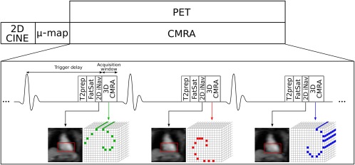
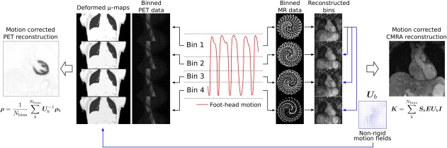
Methods: An acquisition that enables nonrigid motion correction of both CMRA and PET has been developed. The proposed method estimates translational motion from low-resolution 2D MR image navigators acquired at each heartbeat and 3D nonrigid respiratory motion between different respiratory bins from the CMRA data itself. Estimated motion is used for correcting the CMRA as well as the emission and attenuation PET data sets to the same respiratory position. The CMRA approach was studied in 10 healthy subjects and compared for both left and right coronary arteries (LCA, RCA) against a reference scan with diaphragmatic navigator gating and tracking. The PET-CMRA approach was tested in 5 oncology patients with 18 F-FDG myocardial uptake. PET images were compared against uncorrected and gated PET reconstructions.
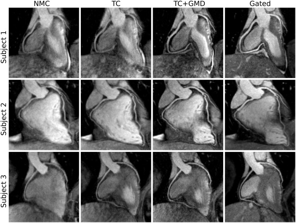
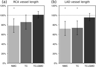
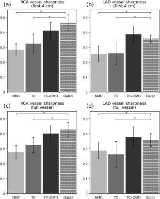
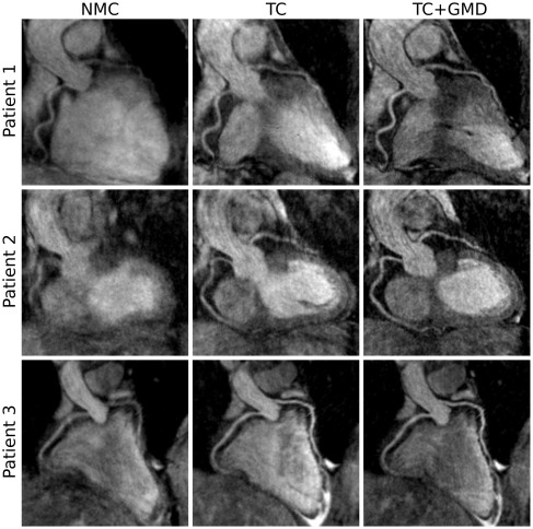
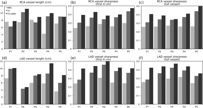
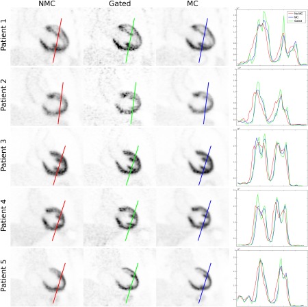
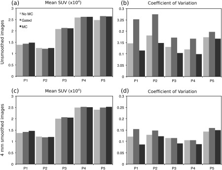
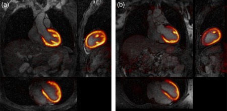
Results: For the healthy subjects, no statistically significant differences in vessel length and sharpness (P > 0.01) were observed between the proposed approach and the reference acquisition with navigator gating and tracking, although data acquisition was significantly shorter. The proposed approach improved CMRA vessel sharpness by 37.9% and 49.1% (LCA, RCA) and vessel length by 48.0% and 36.7% (LCA, RCA) in comparison with no motion correction for all the subjects. Motion-corrected PET images showed improved sharpness of the myocardium compared to uncorrected reconstructions and reduced noise compared to gated reconstructions.
Conclusion: Feasibility of a new respiratory motion-compensated simultaneous cardiac PET-CMRA acquisition has been demonstrated. Magn Reson Med 79:339-350, 2018. © 2017 The Authors Magnetic Resonance in Medicine published by Wiley Periodicals, Inc. on behalf of International Society for Magnetic Resonance in Medicine. This is an open access article under the terms of the Creative Commons Attribution License, which permits use, distribution and reproduction in any medium, provided the original work is properly cited.
Keywords: cardiac PET-MR; coronary MRA; myocardial PET; nonrigid motion correction.
© 2017 The Authors Magnetic Resonance in Medicine published by Wiley Periodicals, Inc. on behalf of International Society for Magnetic Resonance in Medicine.
Figures
References
-
- Karamitsos TD, Francis JM, Myerson S, Selvanayagam JB, Neubauer S. The role of cardiovascular magnetic resonance imaging in heart failure. J Am Coll Cardiol 2009;54:1407–1424. - PubMed
-
- Ratib O, Nkoulou R. Potential applications of PET/MR imaging in cardiology. J Nucl Med 2014;55(Suppl 2):40S–46S. - PubMed
-
- Di Carli MF, Dorbala S, Meserve J, El Fakhri G, Sitek A, Moore SC. Clinical myocardial perfusion PET/CT. J Nucl Med 2007;48:783–793. - PubMed
-
- Rischpler C, Langwieser N, Souvatzoglou M, Batrice A, van Marwick S, Snajberk J, Ibrahim T, Laugwitz KL, Nekolla SG, Schwaiger M. PET/MRI early after myocardial infarction: evaluation of viability with late gadolinium enhancement transmurality vs. 18F‐FDG uptake. Eur Hear J Cardiovasc Imaging 2015;16:661–669. - PubMed
-
- Derlin T, Richter U, Bannas P, Begemann P, Buchert R, Mester J, Klutmann S. Feasibility of 18F‐sodium fluoride PET/CT for imaging of atherosclerotic plaque. J Nucl Med 2010;51:862–865. - PubMed
MeSH terms
Substances
LinkOut - more resources
Full Text Sources
Other Literature Sources
Research Materials
