. 2017 Apr 20;18(1):66.
doi: 10.1186/s12931-017-0551-8.

Monitoring mandibular movements to detect Cheyne-Stokes Breathing

Affiliations

Monitoring mandibular movements to detect Cheyne-Stokes Breathing

Jean-Benoît Martinot et al. Respir Res. .

Abstract

Background: The patterns of mandibular movements (MM) during sleep can be used to identify increased respiratory effort periodic large-amplitude MM (LPM), and cortical arousals associated with "sharp" large-amplitude MM (SPM). We hypothesized that Cheyne Stokes breathing (CSB) may be identified by periodic abnormal MM patterns. The present study aims to evaluate prospectively the concordance between CSB detected by periodic MM and polysomnography (PSG) as gold-standard. The present study aims to evaluate prospectively the concordance between CSB detected by periodic MM and polysomnography (PSG) as gold-standard.

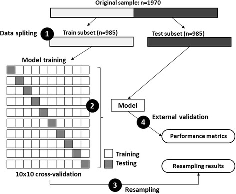
Methods: In 573 consecutive patients attending an in-laboratory PSG for suspected sleep disordered breathing (SDB), MM signals were acquired using magnetometry and scored manually while blinded from the PSG signal. Data analysis aimed to verify the concordance between the CSB identified by PSG and the presence of LPM or SPM. The data were randomly divided into training and validation sets (985 5-min segments/set) and concordance was evaluated using 2 classification models.

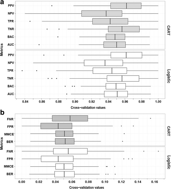
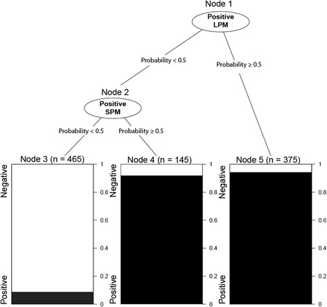
Results: In PSG, 22 patients (mean age ± SD: 65.9 ± 15.0 with a sex ratio M/F of 17/5) had CSB (mean central apnea hourly indice ± SD: 17.5 ± 6.2) from a total of 573 patients with suspected SDB. When tested on independent subset, the classification of CSB based on LPM and SPM is highly accurate (Balanced-accuracy = 0.922, sensitivity = 0.922, specificity = 0.921 and error-rate = 0.078). Logistic models based odds-ratios for CSB in presence of SPM or LPM were 172.43 (95% CI: 88.23-365.04; p < 0.001) and 186.79 (95% CI: 100.48-379.93; p < 0.001), respectively.

Conclusion: CSB in patients with sleep disordered breathing could be accurately identified by a simple magnetometer device recording mandibular movements.

Keywords: Central sleep apnea syndrome; Cheyne Stokes breathing; Polysomnography; Sleep mandibular movements.


Figures

Fig. 1
Fig. 1
Measuring the mandibular movements and definition of periodic MML and MMS. a Forehead sensor, b Chin emitter, d is a distance between emitter and sensor, d0: offset level, Δd: variation of distance d when the mouth opens. CSB: Cheyne-Stokes breathing highlighted by a flow typical crescendo –decrescendo pattern of at least 5 respirations; SPM: periodic sharp mandibular movements occurring on cortical arousals during the hyperventilation phase (these are unevenly observed); LPM: periodic large mandibular movements accompanying the changes in flow during the hyperventilation phase
Fig. 2
Fig. 2
Typical MM recorded during CSB vs Obstructive events for comparing true positive SPM or LPM vs MM during obstructive events. Examples of: a, b Central and c Obstructive respiratory events during a period of 5 sleep minutes. The arrows indicate cortical arousals. SaO2: oxygen saturation; VTH and VAB: thoracic and abdominal inductance belts; NAF2P and NAF1: nasal pressure transducer and oronasal thermal flow sensor; MM: mandibular movements. Cortical arousals are highlighted with an arrow. During the central event (a, b), one issue of classification is presented, including: True Positive for LPM (a) and for SPM (b). During the obstructive event (c), the more negative the signal, the lower the mandibular position and the greater the mouth opening until a sharp and great movement occurs closing the mouth
Fig. 3
Fig. 3
Model training and testing process. (1) data splitting: the original dataset (n = 1970) were randomly divided into two equal sized subsets of fragments (n = 985): one to be used for model training and the other for model testing. (2) the model training process implies a 10 × 10 cross-validation and provides the best fit model. (3) Posterior predictive values of the model were estimated from cross-validation resampling process. (4) Finally, the model was tested against external subset (n = 985). Model’s performance metrics were evaluated
Fig. 4
Fig. 4
Performance of the two models (CART and Logistic) evaluated by cross-validation resampling. a Six metrics for evaluating the model’s performance (Best value = 1): PPV = Positive predictive value or the probability that fragments with positive LPM or SPM truly reflect CSB. NPV = Negative predictive value or the probability that CSB is correctly excluded once neither LPM nor SPM is identified; TPR = True positive rate, or Sensitivity, is the percentage of correctly classified observation among positive CSB class; TNR = True Negative rate or Specificity, is the percentage of correctly excluded CSBs; BAC = Balanced accuracy or Mean of true positive rate and true negative rate; AUC = Area under Receiver Operating Curve (ROC) that results from computing False positive rate and True positive rate from many thresholds. b Four metrics for evaluating the classification error (Best value =0): FNR = False negative rate, or percentage of in the negative CSB class. FPR = False positive rate, or percentage of misclassified observations in the Positive CSB class, MMCE = mean misclassification error, defined as mean of all classifications that disagree with truth; BER = balanced error index, defined as Mean of misclassification error rates on all individual classes
Fig. 5
Fig. 5
Decision tree classifier for detecting CSB by periodic MML and MMS. Decision tree classification rule: First, the model checks whether LPM presents within the segment (Node 1). If so, the segment will be classified as CSB (Node 5). If not, the model will check whether the segment presents a SPM (Node 2). If so, it will be classified as CSB positive (Node 4); if not, CSB will be excluded (Node 3). Based on such rule, absence of both LPM and SPM allows to exclude CSB with an error rate of 8.6% whilst CSB could be ruled in by using either LPM (error rate of 5.9%) or SPM (error rate of 8.3%)

References

    1. Costanzo MR, Khayat R, Ponikowski P, Agostini R, Stellbrink C, Mianulli M, Abraham WT. Mechanisms and clinical consequences of untreated central sleep apnea in heart failure. J Am Coll Cardiol. 2015;65:72–84. doi: 10.1016/j.jacc.2014.10.025. - DOI - PMC - PubMed
    1. Khayat R, Jarjoura D, Porter K, Sow A, Wannemacher J, Dohar R, Pleister A, Abraham WT. Sleep disordered breathing and post-discharge mortality in patients with acute heart failure. Eur Heart J. 2015;36:1463–9. doi: 10.1093/eurheartj/ehu522. - DOI - PMC - PubMed
    1. Matthew T, Naughton T, Kirk K. Sleep apnoea in heart failure: To treat or not to treat? Respirology. 2017;22:217–29. doi: 10.1111/resp.12964. - DOI - PubMed
    1. Hernandez AB, Patil SP. Pathophysiology of central sleep apneas. Sleep Breath. 2016;20:467–82. doi: 10.1007/s11325-015-1290-z. - DOI - PubMed
    1. Leung R, Huber M, Rogge T, Maimon N, Chiu K, Bradley D. Association between atrial fibrillation and central sleep apnea. Sleep. 2005;28:1543–6. doi: 10.1093/sleep/28.12.1543. - DOI - PubMed