. 2017 Apr 20;10(1):193.
doi: 10.1186/s13071-017-2108-6.

Parasites, pathogens and commensals in the "low-impact" non-native amphipod host Gammarus roeselii

Affiliations

Parasites, pathogens and commensals in the "low-impact" non-native amphipod host Gammarus roeselii

Jamie Bojko et al. Parasit Vectors. .

Abstract

Background: Whilst vastly understudied, pathogens of non-native species (NNS) are increasingly recognised as important threats to native wildlife. This study builds upon recent recommendations for improved screening for pathogens in NNS by focusing on populations of Gammarus roeselii in Chojna, north-western Poland. At this location, and in other parts of continental Europe, G. roeselii is considered a well-established and relatively 'low-impact' invader, with little understanding about its underlying pathogen profile and even less on potential spill-over of these pathogens to native species.

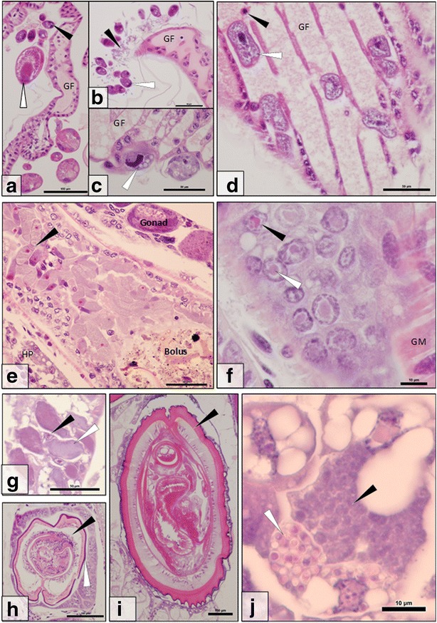
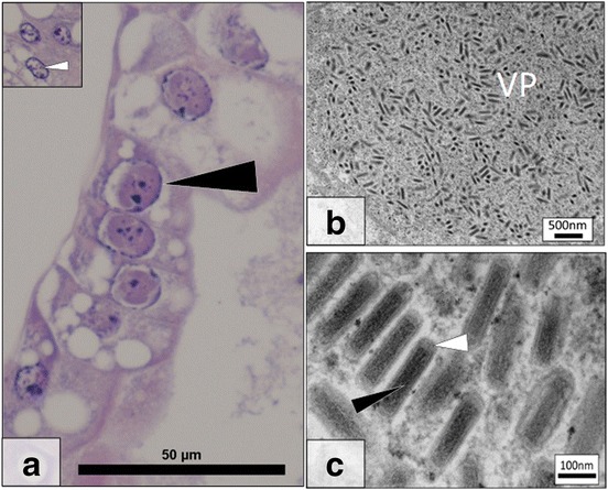
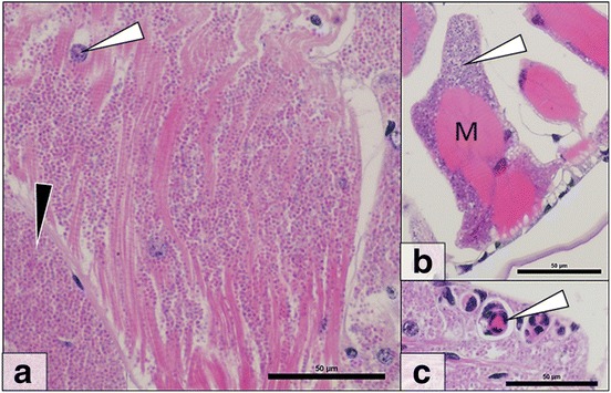
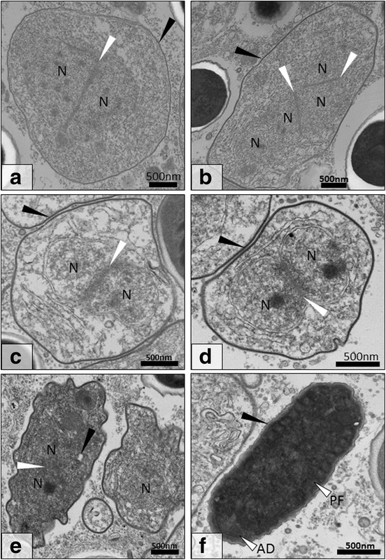
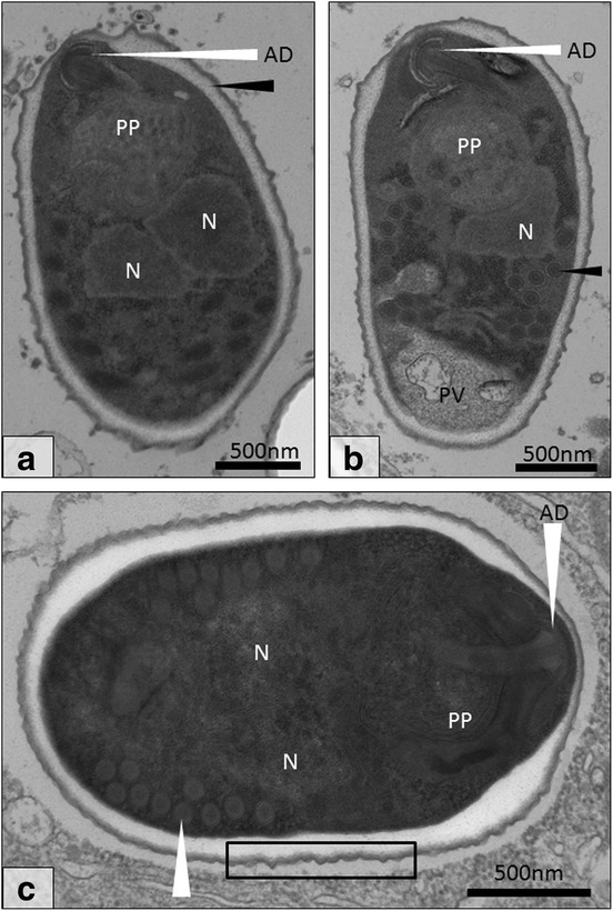
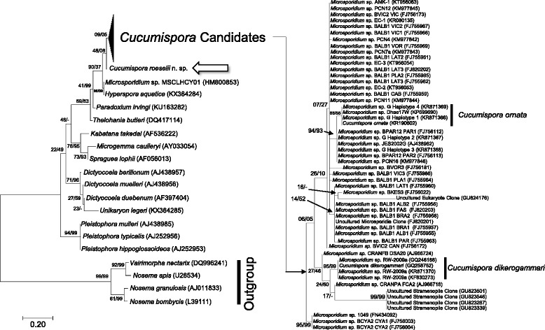
Results: Using a combination of histological, ultrastructural and phylogenetic approaches, we define a pathogen profile for non-native populations of G. roeselii in Poland. This profile comprised acanthocephalans (Polymorphus minutus Goese, 1782 and Pomphorhynchus sp.), digenean trematodes, commensal rotifers, commensal and parasitic ciliated protists, gregarines, microsporidia, a putative rickettsia-like organism, filamentous bacteria and two viral pathogens, the majority of which are previously unknown to science. To demonstrate potential for such pathogenic risks to be characterised from a taxonomic perspective, one of the pathogens, a novel microsporidian, is described based upon its pathology, developmental cycle and SSU rRNA gene phylogeny. The novel microsporidian Cucumispora roeselii n. sp. displayed closest morphological and phylogenetic similarity to two previously described taxa, Cucumispora dikerogammari (Ovcharenko & Kurandina, 1987), and Cucumispora ornata Bojko, Dunn, Stebbing, Ross, Kerr & Stentiford, 2015.

Conclusions: In addition to our discovery extending the host range for the genus Cucumispora Ovcharenko, Bacela, Wilkinson, Ironside, Rigaud & Wattier, 2010 outside of the amphipod host genus Dikerogammarus Stebbing, we reveal significant potential for the co-transfer of (previously unknown) pathogens alongside this host when invading novel locations. This study highlights the importance of pre-invasion screening of low-impact NNS and, provides a means to document and potentially mitigate the additional risks posed by previously unknown pathogens.

Keywords: Amphipoda; Cucumispora; Invasive; Microsporidia; Parasite; Virus; Wildlife disease.


Figures

Fig. 1
Fig. 1
Parasites and pathogens observed during the histological screen of Gammarus roeselii. a External rotifers (white arrow) and stalked ciliated protists (black arrow) clustered around a gill filament (GF). b Externally associated ciliated protists (white arrow) and filamentous bacteria (black arrow) clustered around a gill filament (GF). c Ciliated protists (white arrow) embedded into the gill filament (GF). d Ciliated protists (white arrow) present in the blood stream (blood cell = black arrow) of the gill filament (GF). e Dense cluster of gregarines (black arrow) in the gut alongside bolus, gonad and hepatopancreas (HP). f Putative nuclei-targeted gut epithelia virus displaying nuclear hypertrophy due to expanding viroplasm (arrows) (GM = gut muscle). g Putative rickettsia-like organism in the cytoplasm of hepatopancreatocytes (white arrow). The nucleus (black arrow) is unaffected. h Digenean trematode (black arrow), present with external pearling (white arrow), encysted internally within G. roeselii. i Polymorphus sp. encysted internally within G. roeselii. j An unidentified microsporidian pathogen in the cytoplasm of infected hepatopancreatocytes. Developing (black arrow) and mature spore stages (white arrow) of the pathogen can be clearly identified in separate cells. Scale-bars: a, h, i, 100 μm; b-e, g, 50 μm; f, j, 10 μm
Fig. 2
Fig. 2
Gammarus roeselii Bacilliform Virus (GrBV) histopathology and ultrastructure. a Several virally infected, hypertrophic, nuclei (black arrow) in the hepatopancreas. Inset at the same magnification details a cluster of uninfected nuclei (white arrow). b Electron micrograph detailing a growing viroplasm (VP) in a nucleus of the hepatopancreas. c High magnification image of the bacilliform virus present with electron dense core (black arrow) and membrane (white arrow) in a paracrystalline array within a heavily infected cell nucleus. Scale-bars: a, 50 μm; b, 500 nm; c, 100 nm
Fig. 3
Fig. 3
Cucumispora roeselii n. sp. histopathology. a Microsporidian spores (black arrow) can be seen throughout the musculature in heavy infections. Muscle nuclei (white arrow) can be seen amongst parasite spores. b Early stage microsporidian infected muscle blocks (M) demonstrate initial sarcolemma infection (white arrow). c Immune reactions (white arrow) towards microsporidian infection. Scale-bars: 50 μm
Fig. 4
Fig. 4
Transmission electron micrograph of early spore development for Cucumispora roeselii n. sp. a Diplokaryotic meront displaying attached nuclei (N; white arrow). Note the thin cell membrane (black arrow). b Tetranucleate cell displaying four attached nuclei (N; white arrows) with a thickening cell wall (black arrow). c After division, two early diplokaryotic (N; white arrow) sporoblasts are produced with further cell membrane thickening (black arrow). d Early diplokaryotic (N; white arrow) sporoblast displaying further thickening of the cell membrane (black arrow). e The early sporoblast begins to become electron dense and condense with some early development of spore organelles such as the polar filament (black arrow). f Fully condensed sporoblast development stage present with electron dense cytoplasm and coiled polar filament (PF) and anchoring disk (AD). At this stage the formation of the early endospore is visible (white arrow). Scale-bars: 500 nm
Fig. 5
Fig. 5
Final development stages of Cucumispora roeselii n. sp. a Diplokaryotic sporoblast (N) with anchoring disk (AD), polaroplast (PP) and thickened endospore (black arrow). b A second sporoblast displaying a clear polar vacuole (PV) and polar filament with rings of varying electron density (black arrow). c The final diplokaryotic (N) spore with bilaminar polaroplast (PP), anchoring disk (AD) and polar filament (9–10 turns; white arrow). The spore wall thins at the anchoring disk (AD) whilst being thickest at the periphery of the anchoring disk. Note the ‘thorned’ spore exterior (black rectangle). Scale-bars: 500 nm
Fig. 6
Fig. 6
A maximum likelihood tree including the bootstrap confidence for ML/NJ phylogenies. If the neighbour joining phylogeny did not produce a node observed on the maximum likelihood tree, a ‘-’ is noted. The tree is displaying the position of Cucumispora roeselii n. sp. (arrow), Cucumispora-related SSU isolates (“Cucumispora Candidates”), various ‘Clade V’ representatives, and various ‘Clade IV’ representatives (as according to Vossbrinck & Debrunner-Vossbrinck [42] as a fungal outgroup. Sequences considered to belong to existing members of the Cucumispora are labelled with the scientific name and indicated by black bars

References

    1. Dunn AM, Hatcher MJ. Parasites and biological invasions: parallels, interactions, and control. Trends Parasitol. 2015;31(5):189–199. doi: 10.1016/j.pt.2014.12.003. - DOI - PubMed
    1. Roy HE, Hesketh H, Purse BV, Eilenberg J, Santini A, Scalera R, et al. Alien pathogens on the horizon: Opportunities for predicting their threat to wildlife. Conserv Lett. 2016;0(0):1–8.
    1. Bojko J, Stebbing PD, Bateman KS, Meatyard JE, Bacela-spychalska K, Dunn AM, et al. Baseline histopathological survey of a recently invading island population of ‘killer shrimp’, Dikerogammarus villosus. Dis Aquat Org. 2013;106(3):241–253. doi: 10.3354/dao02658. - DOI - PubMed
    1. Bij de Vaate A, Jazdzewski K, Ketelaars HA, Gollasch S, Van der Velde G. Geographical patterns in range extension of Ponto-Caspian macroinvertebrate species in Europe. Can J Fish Aquat Sci. 2002;59(7):1159–1174. doi: 10.1139/f02-098. - DOI
    1. Grabowski M, Jazdzewski K, Konopacka A. Alien Crustacea in Polish waters - Amphipoda. Aquat Invasions. 2007;2(1):25–38. doi: 10.3391/ai.2007.2.1.3. - DOI

MeSH terms

LinkOut - more resources