Multicenter Study
. 2017 Apr 24;10(8):798-809.
doi: 10.1016/j.jcin.2017.01.035.

Intentional Percutaneous Laceration of the Anterior Mitral Leaflet to Prevent Outflow Obstruction During Transcatheter Mitral Valve Replacement: First-in-Human Experience

Affiliations
Multicenter Study

Intentional Percutaneous Laceration of the Anterior Mitral Leaflet to Prevent Outflow Obstruction During Transcatheter Mitral Valve Replacement: First-in-Human Experience

Vasilis C Babaliaros et al. JACC Cardiovasc Interv. .

Abstract

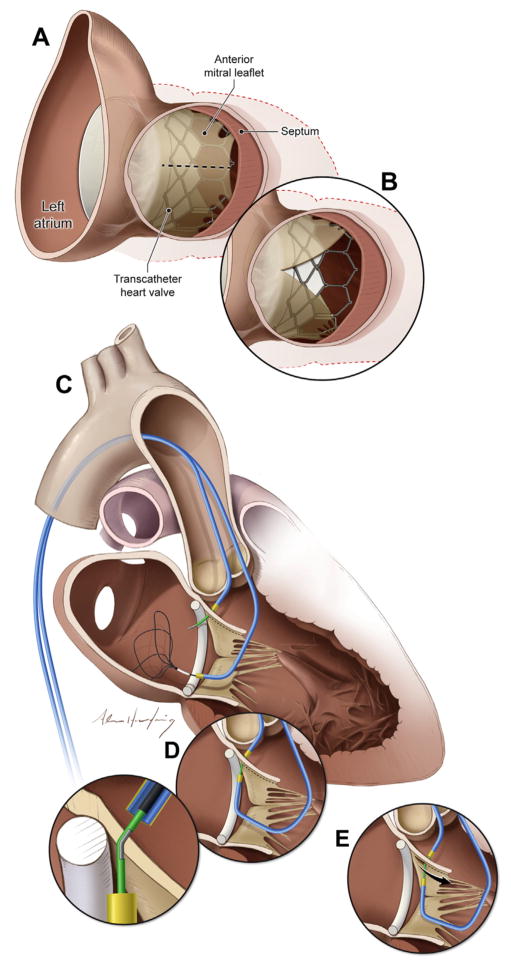
Objectives: This study sought to use a new catheter technique to split the anterior mitral valve leaflet (AML) and prevent iatrogenic left ventricular outflow tract (LVOT) obstruction immediately before transcatheter mitral valve replacement (TMVR).

Background: LVOT obstruction is a life-threatening complication of TMVR, caused by septal displacement of the AML.

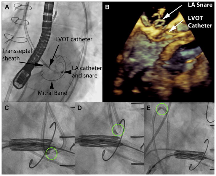
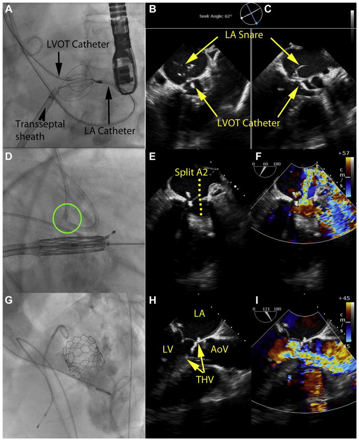
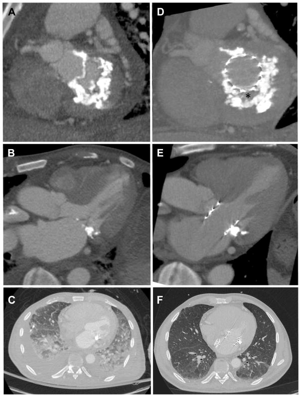
Methods: The procedure was used in patients with severe mitral valve disease and prohibitive surgical risk. Patients either had prior surgical mitral valve ring (n = 3) or band annuloplasty (n = 1) or mitral annular calcification with stenosis (n = 1). Iatrogenic LVOT obstruction or transcatheter heart valve dysfunction was predicted in all based on echocardiography and computed tomography. Transfemoral coronary guiding catheters directed an electrified guidewire across the center and base of the AML toward a snare in the left atrium. The externalized guidewire loop was then electrified to lacerate the AML along the centerline from base to tip, sparing chordae, immediately before transseptal TMVR.

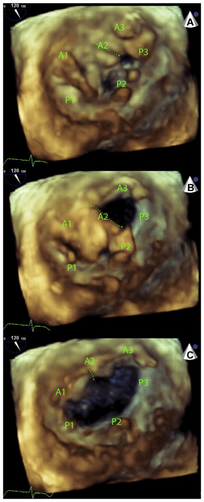
Results: Five patients with prohibitive risk of LVOT obstruction or transcatheter heart valve dysfunction from TMVR successfully underwent LAMPOON, with longitudinal splitting of the A2 scallop of the AML, before valve implantation. Multiplane computed tomography modeling predicted hemodynamic collapse from TMVR assuming an intact AML. However, critical LVOT gradients were not seen following LAMPOON and TMVR. Doppler blood flow was seen across transcatheter heart valve struts that encroached the LVOT, because the AML was split. Transcatheter heart valve function was unimpeded.

Conclusions: This novel catheter technique, which resembles surgical chord-sparing AML resection, may enable TMVR in patients with prohibitive risk of LVOT obstruction or transcatheter heart valve dysfunction.

Keywords: left ventricular outflow tract obstruction; mitral valve; structural heart disease; transcatheter mitral valve replacement; valvular heart disease.


Figures

FIGURE 1
FIGURE 1. Views of the Anterior Mitral Valve Leaflet From the LVOT
(A) In this example, transcatheter mitral valve implantation displaces the native anterior mitral valve leaflet causing LVOT obstruction. (B) After LAMPOON, LVOT obstruction is reduced and blood flows across the unobstructed struts of the implanted transcatheter heart valve. (C to E) Illustration of the LAMPOON procedure. (C) In 2 retrograde guiding catheters are positioned across the aortic valve, 1 into the LVOT and another across the mitral valve into the left atrium. The LVOT catheter directs an electrified guidewire across the base of the anterior mitral valve leaflet under echocardiographic guidance into a snare positioned through the left atrial guiding catheter. (D) Once the guidewire traverses the mitral leaflet, it is ensnared and externalized. The inset depicts a denuded and kinked section of the guidewire shaft that directs electrosurgery energy to the leaflet. (E) The guidewire is electrified and the 2 guiding catheters withdraw it to lacerate the anterior mitral leaflet lengthwise. LAMPOON = laceration of the anterior mitral leaflet to prevent left ventricular outflow tract obstruction; LVOT = left ventricular outflow tract. Online Video 1 is depicted in this figure.
FIGURE 2
FIGURE 2. LAMPOON to Enable Transcatheter Mitral Valve Replacement Inside a Flexible Mitral Annuloplasty Band
(A) Left anterior oblique caudal short-axis fluoroscopic projection showing 2 retrograde catheters positioned before anterior mitral valve leaflet traversal. The retrograde LVOT and LA catheters overlap in this view, as intended. The posterior mitral valve band provides a fluoroscopic marker to position the LVOT catheter along the base of the A2 scallop. The LA catheter is directing the multiloop snare and is supported by a transseptal rail through a transseptal sheath. (B) A 3-dimensional transesophageal echocardiograph of the same step, with a LVOT catheter positioned at the base of the A2 scallop, and the LA catheter pointing a multiloop snare at the other side of the A2 scallop. (C) Initiation of laceration. The kinked guidewire cutting edge is circled in green. The transcatheter heart valve is pre-positioned at the orifice of the mitral valve. The LAMPOON guidewire is pulled outward to apply tension. (D) The LAMPOON guidewire is electrified during further pulling to initiate laceration. (E) Completed laceration, with both retrograde catheters insulating the wire safely in the descending aorta. The kinked guidewire cutting edge, adjacent to the radiopaque piggyback tip marker, is seen sheathed in the catheter. LA = left atrium; other abbreviation as in Figure 1. Online Video 2 is depicted in this figure.
FIGURE 3
FIGURE 3. Imaging Immediately After LAMPOON for TMVR-in-Band
These images are from the same patient as in Figure 2. (A) B-mode and color Doppler transesophageal echocardiography after LAMPOON and TMVR shows the stent struts protruding into the LVOT, and blood flow across those struts indicating successful leaflet splitting. (B) Catheter pressure measurements across the LVOT show a tolerable gradient of 16 mm Hg. (C) Left ventriculography and (D) contrast computed tomography after TMVR shows the THV encroaches completely across the LVOT and would nearly completely obstruct it if not for LAMPOON. AoV = aortic valve; LV = left ventricle; THV = transcatheter heart valve; TMVR = transcatheter mitral valve replacement; other abbreviations as in Figures 1 and 2. Online Video 2 is depicted in this figure.
FIGURE 4
FIGURE 4. LAMPOON to Enable TMVR in Native Mitral Annular Calcification
(A to C) Leaflet traversal, (D to F) leaflet laceration, and (G to I) imaging after LAMPOON and TMVR. (A) Guidewire traversal across the base of the anterior mitral leaflet. Four catheters are in place. An antegrade transseptal sheath, used for TMVR, is currently connected via a guidewire rail to control the retrograde LA catheter, which is used to deliver the multiloop snare on the LA side of the anterior mitral leaflet. A retrograde catheter in the LVOT is directing the traversing guidewire. There also is a pigtail catheter in the ascending aorta. (B, C) X-plane TEE immediately after leaflet traversal shows the mid-basal A2 position of the traversal system. Also evident are the 2 catheters and the LA snare. (D) The traversing guidewire tip has been externalized. The guidewire shaft is denuded, kinked, and exposed between the 2 catheters (green circle), and is electrified to slice the anterior mitral leaflet longitudinally. The THV is prepositioned for immediate deployment. (E, F) B-mode and color Doppler TEE after LAMPOON but before TMVR shows severe acute mitral valve regurgitation across the split A2 leaflet scallop. (G) Left ventriculography after TMVR shows the THV encroaches nearly completely across the LVOT and would nearly completely obstruct it if not for LAMPOON. (H, I) B-mode and color Doppler TEE after LAMPOON and TMVR shows the stent struts protruding into the LVOT, and blood flow across those struts indicating successful leaflet splitting. TEE = transesophageal echocardiography; other abbreviations as in Figures 1 to 3.
FIGURE 5
FIGURE 5. LAMPOON-Induced Anterior Mitral Leaflet Split
Three sequential diastolic frames of a 3-dimensional echocardiogram performed immediately after LAMPOON but before TMVR. The volume is rendered from a left-atrial “surgeons view.” (A) A laceration cleft is evident (dotted green line) at the beginning of diastole in the A2 scallop of the anterior mitral leaflet. (B) The medial half of A2 and the whole of A3 open early in diastole and then (C) the lateral half of A2 along with the whole of A1 open a fraction later in diastole. A1 to A3 and P1 to P3 represent the lateral to medial scallops of the anterior and posterior mitral leaflets, respectively. Abbreviations as in Figures 1 and 3.
FIGURE 6
FIGURE 6. LAMPOON and TMVR in Mitral Annular Calcification
Computed tomography scans before (A to C) and after (D to F) TMVR with LAMPOON in a patient treated for mitral annular calcification causing mitral valve stenosis. Noncalcified fibrous valve tissue (asterisk) is interposed between the THV and the mitral annular calcification (D). The transcatheter heart valve is shown to span across the LVOT (E), suggesting there would have been complete LVOT obstruction had LAMPOON not been performed. Follow-up chest images (F) show near-resolution of pulmonary alveolar hemorrhage and pleural effusion. Abbreviations as in Figures 1 and 3.

References

    1. Anderson RH, Spicer DE, Hlavacek AM, Cook AC, Backer CL. Wilcox’s Surgical Anatomy of the Heart. 4. New York: Cambridge University Press; 2013.
    1. Come PC, Riley MF, Weintraub RM, et al. Dynamic left ventricular outflow tract obstruction when the anterior leaflet is retained at prosthetic mitral valve replacement. Ann Thorac Surg. 1987;43:561–3. - PubMed
    1. Esper E, Ferdinand FD, Aronson S, Karp RB. Prosthetic mitral valve replacement: late complications after native valve preservation. Ann Thorac Surg. 1997;63:541–3. - PubMed
    1. Bapat V, Pirone F, Kapetanakis S, Rajani R, Niederer S. Factors influencing left ventricular outflow tract obstruction following a mitral valve-in-valve or valve-in-ring procedure, part 1. Catheter Cardiovasc Interv. 2015;86:747–60. - PubMed
    1. Hamid NB, Khalique OK, Monaghan MJ, et al. Transcatheter valve implantation in failed surgically inserted bioprosthesis: review and practical guide to echocardiographic imaging in valve-in-valve procedures. J Am Coll Cardiol Img. 2015;8:960–79. - PubMed

Publication types

MeSH terms