. 2017 Jun;187(6):1368-1379.
doi: 10.1016/j.ajpath.2017.02.007. Epub 2017 Apr 17.

Divergent Function of Programmed Death-Ligand 1 in Donor Tissue versus Recipient Immune System in a Murine Model of Bronchiolitis Obliterans

Affiliations

Divergent Function of Programmed Death-Ligand 1 in Donor Tissue versus Recipient Immune System in a Murine Model of Bronchiolitis Obliterans

Katharina Schütte-Nütgen et al. Am J Pathol. 2017 Jun.

Abstract

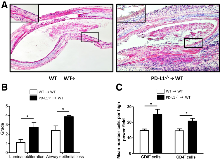
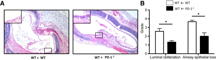
Costimulatory molecules, such as the programmed death ligand (PD-L1), might exert differential effects on T-cell function, depending on the clinical setting and/or immunological environment. Given the impact of T cells on bronchiolitis obliterans (BO) in lung transplantation, we used an established tracheal transplant model inducing BO-like lesions to investigate the impact of PD-L1 on alloimmune responses and histopathological outcome in BO. In contrast to other transplant models in which PD-L1 generally shows protective functions, we demonstrated that PD-L1 has divergent effects depending on its location in donor versus recipient tissue. Although PD-L1 deficiency in donor tissue worsened histopathological outcome, and increased systemic inflammatory response, recipient PD-L1 deficiency induced opposite effects. Mechanistic studies revealed PD-L1-deficient recipients were hyporesponsive toward alloantigen, despite increased numbers of CD8+ effector T cells. The function of PD-L1 on T cells after unspecific stimulation was dependent on both cell type and strength of stimulation. This novel function of recipient PD-L1 may result from the high degree of T-cell activation within the highly immunogenic milieu of the transplanted tissue. In this model, both decreased T-cell alloimmune responses and the reduction of BO in PD-L1-deficient recipients suggest a potential therapeutic role of selectively blocking PD-L1 in the recipient. Further investigation is warranted to determine the impact of this finding embedded in the complex pathophysiological context of BO.


Figures

Figure 1
Figure 1
PD-L1 is up-regulated in airway epithelial cells of tracheal allografts. A: Frozen sections of naïve tracheas and tracheal allografts at day 14 after transplantation were stained with an antibody to PD-L1 (immunohistochemistry). PD-L1 expression is low in the epithelium of naive BALB/c tracheas but up-regulated in BALB/c tracheal allografts 14 days after transplantation into B6 recipients. Boxed areas indicate the tracheal epithelium and are shown at higher magnification in the insets. B: Flow cytometry was performed on tracheal tissue and CD45CD326+ cells were identified as epithelial cells. PD-L1 expression in epithelial cells was measured as fluorescence intensity. Tracheal allografts show up-regulation of PD-L1 on epithelial cells on day 14 after transplantation compared to naïve tracheas. Original magnification, ×10 (A). FMO, fluorescence minus one control.
Figure 2
Figure 2
PD-L1–deficient donor grafts are more susceptible to BO development. Tracheas from WT and PD-L1−/− B6 donors were transplanted into WT BALB/c recipients and harvested 14 days after transplantation. A: Cross sections of paraffin-embedded tracheal tissues (5 μm thick) were stained with hematoxylin and eosin to assess the degree of luminal obliteration and epithelial loss. Boxed areas indicate the tracheal epithelium and are shown at higher magnification in the insets. B: Sections were graded by two independent investigators (K.S.-N., M.Su.) who were blinded for the groups. PD-L1−/− donor tracheas show more severe luminal obliteration and airway epithelial loss compared to WT grafts. C: Immunohistochemistry analysis of allograft infiltrating CD4+ and CD8+ T cells was performed on frozen tracheal sections and reveals significantly higher numbers in PD-L1−/− allografts when compared to WT allografts. Cells were counted in five random high-power fields. Data are representative of three transplant series. n = 4 animals per group (AC). P < 0.05. Original magnification, ×10 (A).
Figure 3
Figure 3
PD-L1–deficient recipients are protected from BO. Tracheas from WT BALB/c donors were transplanted into WT and PD-L1−/− B6 recipients and harvested 14 days after transplantation. A: Cross sections of paraffin-embedded tracheal tissues (5 μm thick) were stained with hematoxylin and eosin to assess the degree of luminal obliteration and epithelial loss. Boxed areas indicate the tracheal epithelium and are shown at higher magnification in the insets. B: Sections were graded by two independent investigators (K.S.-N., M.Su.) who were blinded for the groups. PD-L1−/− recipients show significantly decreased degrees of luminal obliteration and loss of airway epithelium. C: Immunohistochemical analysis of allograft infiltrating CD4+ and CD8+ T cells was performed on frozen tracheal sections and reveals significantly higher numbers in PD-L1−/− recipients, when compared to WT recipients. Cells were counted in five random high-power fields. Data are representative of four transplant series. n = 4 animals at least per group (AC). P < 0.05, ∗∗∗P < 0.001. Original magnification, ×10 (A).
Figure 4
Figure 4
PD-1–deficient recipients are protected from BO. Tracheas from WT BALB/c donors were transplanted into WT and PD-1−/− B6 recipients and harvested 14 days after transplantation. A: Cross sections of paraffin-embedded tracheal tissues (5 μm thick) were stained with hematoxylin and eosin to assess the degree of luminal obliteration and epithelial loss. Boxed areas indicate the tracheal epithelium and are shown at higher magnification in the insets. B: Sections were graded by two independent investigators (K.S.-N., M.Su.) who were blinded for the groups. Histopathological grading reveals significantly reduced degrees of luminal obliteration and loss of airway epithelium in PD-1−/− B6 recipients compared to WT B6 recipients. Data are representative of four transplant series. n = 4 animals at least per group (A and B). P < 0.05. Original magnification, ×10 (A).
Figure 5
Figure 5
Overexpression of PD-1 on CD4+ and CD8+ effector T cells is not required for graft protection in PD-L1–deficient recipients. A: Tracheas from WT BALB/c donors were transplanted into WT and PD-L1−/− B6 recipients, and spleens were harvested 7 days after transplantation. PD-1 expression on T-effector cells was analyzed by flow cytometry. The percentage of PD-1–positive CD4+ and CD8+ effector cells is significantly higher in PD-L1−/− recipients. Data are representative of four different experiments. B: To test whether the enhanced PD-1 expression and a subsequent increased interaction between PD-1 on PD-L1−/− immune cells and PD-L1 within the graft tissue mediates the protective effects observed in PD-L1−/− recipients, PD-1 was blocked in these mice by administration of a monoclonal antibody against PD-1. Cross sections of paraffin-embedded tracheal tissues (5 μm thick) were stained with hematoxylin and eosin to assess the degree of luminal obliteration and epithelial loss. Sections were graded by two independent investigators (K.S.-N., M.Su.) who were blinded for the groups. Histopathological scoring did not reveal any significant effects of PD-1 blockade in PD-L1−/− recipients when compared to isotype-treated controls 14 days after transplantation. Data are representative of three different experiments. n = 4 animals per group (A and B). P < 0.05, ∗∗∗P < 0.001.
Figure 6
Figure 6
Recipients of PD-L1–deficient tracheal allografts show increased systemic alloimmune responses. Tracheas from WT and PD-L1−/− B6 donors were transplanted into WT BALB/c recipients. To assess systemic alloimmune response in the recipient, spleens were harvested 7 days after transplantation. A: Flow cytometry analysis of CD4+ and CD8+ effector cells (CD44highCD62Llow phenotype) of spleens from WT recipients of PD-L1−/− tracheas show increased percentages of CD4+ and CD8+ effector cells when compared to WT recipients of WT tracheas 7 days after transplantation. Frequencies of effector and regulatory T cells are given as percentage of the total CD4+ or CD8+ population. B: Similarly, the frequencies of cytokine-producing splenocytes, as analyzed by ELISPOT assays, increase in WT recipients of PD-L1−/− tracheas 7 days after transplantation. Frequencies are expressed as the number of spots per 0.5 × 106 responder cells. ELISPOT experiments were performed in triplicate. Data are representative of three independent experiments. n = 5 animals at least per group (A and B). P < 0.05, ∗∗P < 0.01, and ∗∗∗P < 0.001.
Figure 7
Figure 7
PD-L1–deficient recipients show decreased systemic alloimmune responses. Tracheas from WT BALB/c donors were transplanted into WT and PD-L1−/− B6 recipients. To assess the systemic alloimmune response in the recipient, spleens were harvested 7 days after transplantation. A: Flow cytometry analysis of CD4+ and CD8+ effector cells (CD44highCD62Llow phenotype) and CD4+ T regulatory cells (Tregs; CD4+CD25+FoxP3+) of spleens from PD-L1−/− recipients shows increased percentages of CD4+ and CD8+ effector cells and CD4+ Tregs when compared to WT recipients 7 days after transplantation. Frequencies of effector and regulatory T cells are given as percentage of the total CD4+ or CD8+ population. B: Despite the increased numbers of effector T cells, PD-L1−/− recipients show decreased frequencies of cytokine-producing splenocytes 7 days after transplantation compared to WT recipients, as assessed by ELSIPOT assays. Frequencies are expressed as the number of spots per 0.5 × 106 responder cells. ELISPOT experiments were performed in triplicate. Data are representative of four independent experiments. n = 4 animals per group (A and B). P < 0.05, ∗∗P < 0.01, and ∗∗∗P < 0.001.
Figure 8
Figure 8
PD-1–deficient recipients show decreased systemic alloimmune responses. Tracheas from WT BALB/c donors were transplanted into WT and PD-1−/− B6 recipients or PD-L1−/−B6 recipients treated with an anti–PD-1 antibody. To assess the systemic alloimmune response in the recipient, spleens were harvested 7 days after transplantation. A: ELISPOT analysis shows significantly decreased frequencies of cytokine-producing splenocytes in PD-1−/− B6 recipients of BALB/c tracheas 7 days after transplantation. B: PD-1 blockade in PD-L1−/− recipients does not affect systemic alloimmune response. ELISPOT assays show similar frequencies of cytokine-producing splenocytes with and without PD-1 blockade (shown for IFN-γ only). Frequencies are expressed as the number of spots per 0.5 × 106 responder cells. ELISPOT experiments were performed in triplicate. Data are representative of four different experiments. n = 4 animals per group (A and B). P < 0.05, ∗∗P < 0.01, and ∗∗∗P < 0.001.
Figure 9
Figure 9
The function of PD-L1 depends on the cell type and the degree of T-cell stimulation. A: Tracheas from WT BALB/c donors were transplanted into WT and PD-1−/− B6 recipients. Flow-sorted CD4+ and CD8+ effector T cells (CD44highCD62Llow phenotype) from WT or PD-L1−/−recipients at day 7 after transplantation were restimulated with donor antigen in an IFN-γ ELISPOT assay. CD4+ T-cell response is low generally in both WT and PD-L1−/− recipients, whereas CD8+ effector cells from WT recipients show notably increased IFN-γ production on restimulation with alloantigen. In contrast, CD8+ effector T cells from PD-L1−/− recipients are hyporesponsive toward alloantigen. Frequencies are expressed as the number of spots per 0.5 × 106 responder cells. B: CD4+ cells from naïve WT and PD-L1−/− B6 animals were isolated by magnetic cell separation and stimulated with low and high concentrations of plate-bound anti-CD3 and soluble anti-CD28. Measurements of IFN-γ production by Luminex assays shows that the regulatory effect of PD-L1 on the activation of CD4+ T cells is restricted to low levels of T-cell activation, whereas it is obscured at high levels of T-cell activation. C: CD8+ cells from naïve WT and PD-L1−/− B6 animals were isolated by magnetic cell separation and stimulated with low and high concentrations of plate-bound anti-CD3 and soluble anti-CD28. In PD-L1−/− CD8+ T cells, IFN-γ production is generally inhibited, regardless of the degree of T-cell stimulation (Luminex). ELISPOT and Luminex experiments were performed in triplicate. Data are representative of three different experiments. n = 4 animals per group (AC). P < 0.05, ∗∗P < 0.01, and ∗∗∗P < 0.001.

Similar articles

Cited by

References

    1. Li X.C., Rothstein D.M., Sayegh M.H. PD-L1 regulates the development, maintenance, and function of induced regulatory T cells. Immunol Rev. 2009;229:271–293. - PubMed
    1. Alegre M.L., Najafian N. Costimulatory molecules as targets for the induction of transplantation tolerance. Curr Mol Med. 2006;6:843–857. - PubMed
    1. Francisco L.M., Salinas V.H., Brown K.E., Vanguri V.K., Freeman G.J., Kuchroo V.K., Sharpe A.H. PD-L1 regulates the development, maintenance, and function of induced regulatory T cells. J Exp Med. 2009;206:3015–3029. - PMC - PubMed
    1. Greenwald R.J., Freeman G.J., Sharpe A.H. The B7 family revisited. Annu Rev Immunol. 2005;23:515–548. - PubMed
    1. Chen L. Co-inhibitory molecules of the B7-CD28 family in the control of T-cell immunity. Nat Rev Immunol. 2004;4:336–347. - PubMed

MeSH terms