

Force development and intracellular Ca2+ in intact cardiac muscles from gravin mutant mice
- PMID: 28428008
- PMCID: PMC5490489
- DOI: 10.1016/j.ejphar.2017.04.020
Force development and intracellular Ca2+ in intact cardiac muscles from gravin mutant mice
Abstract
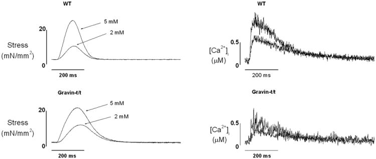
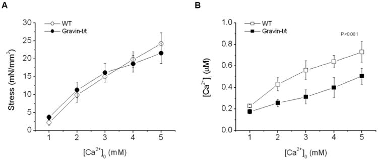
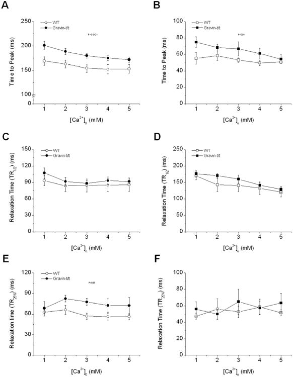
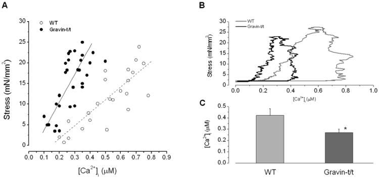
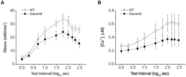
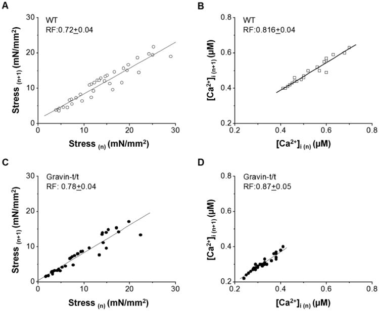
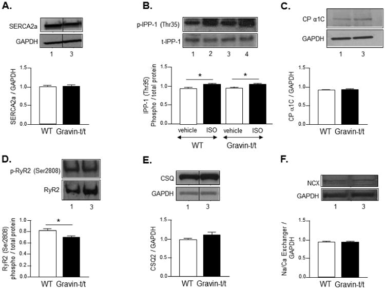
Gravin (AKAP12) is an A-kinase-anchoring-protein that scaffolds protein kinase A (PKA), β2-adrenergic receptor (β2-AR), protein phosphatase 2B and protein kinase C. Gravin facilitates β2-AR-dependent signal transduction through PKA to modulate cardiac excitation-contraction coupling and its removal positively affects cardiac contraction. Trabeculae from the right ventricles of gravin mutant (gravin-t/t) mice were employed for force determination. Simultaneously, corresponding intracellular Ca2+ transient ([Ca2+]i) were measured. Twitch force (Tf)-interval relationship, [Ca2+]i-interval relationship, and the rate of decay of post-extrasysolic potentiation (Rf) were also obtained. Western blot analysis were performed to correlate sarcomeric protein expression with alterations in calcium cycling between the WT and gravin-t/t hearts. Gravin-t/t muscles had similar developed force compared to WT muscles despite having lower [Ca2+]i at any given external Ca2+ concentration ([Ca2+]o). The time to peak force and peak [Ca2+]i were slower and the time to 75% relaxation was significantly prolonged in gravin-t/t muscles. Both Tf-interval and [Ca2+]i-interval relations were depressed in gravin-t/t muscles. Rf, however, did not change. Furthermore, Western blot analysis revealed decreased ryanodine receptor (RyR2) phosphorylation in gravin-t/t hearts. Gravin-t/t cardiac muscle exhibits increased force development in responsiveness to Ca2+. The Ca2+ cycling across the SR appears to be unaltered in gravin-t/t muscle. Our study suggests that gravin is an important component of cardiac contraction regulation via increasing myofilament sensitivity to calcium. Further elucidation of the mechanism can provide insights to role of gravin if any in the pathophysiology of impaired contractility.
Keywords: Cardiac muscle; Contraction; Force development; Gravin; Intracellular calcium; Trabeculae.
Copyright © 2017 Elsevier B.V. All rights reserved.
Figures
Similar articles
-
Enhanced cardiac function in Gravin mutant mice involves alterations in the β-adrenergic receptor signaling cascade.PLoS One. 2013 Sep 18;8(9):e74784. doi: 10.1371/journal.pone.0074784. eCollection 2013. PLoS One. 2013. PMID: 24058627 Free PMC article.
-
Receptor-mediated Ca2+ and PKC signaling triggers the loss of cortical PKA compartmentalization through the redistribution of gravin.Cell Signal. 2013 Nov;25(11):2125-35. doi: 10.1016/j.cellsig.2013.07.004. Epub 2013 Jul 6. Cell Signal. 2013. PMID: 23838009 Free PMC article.
-
High-mobility group box 1 (HMGB1) impaired cardiac excitation-contraction coupling by enhancing the sarcoplasmic reticulum (SR) Ca(2+) leak through TLR4-ROS signaling in cardiomyocytes.J Mol Cell Cardiol. 2014 Sep;74:260-73. doi: 10.1016/j.yjmcc.2014.06.003. Epub 2014 Jun 14. J Mol Cell Cardiol. 2014. PMID: 24937603
-
β-Adrenergic modulation of skeletal muscle contraction: key role of excitation-contraction coupling.J Physiol. 2015 Nov 1;593(21):4713-27. doi: 10.1113/JP270909. J Physiol. 2015. PMID: 26400207 Free PMC article. Review.
-
[Excitation-Contraction coupling and intracellular calcium cycling in failing hearts].Clin Calcium. 2013 Apr;23(4):471-80. Clin Calcium. 2013. PMID: 23545736 Review. Japanese.
Cited by
-
Human muscle-specific A-kinase anchoring protein polymorphisms modulate the susceptibility to cardiovascular diseases by altering cAMP/PKA signaling.Am J Physiol Heart Circ Physiol. 2018 Jul 1;315(1):H109-H121. doi: 10.1152/ajpheart.00034.2018. Epub 2018 Mar 30. Am J Physiol Heart Circ Physiol. 2018. PMID: 29600899 Free PMC article.
-
Nanodomain cAMP signaling in cardiac pathophysiology: potential for developing targeted therapeutic interventions.Physiol Rev. 2025 Apr 1;105(2):541-591. doi: 10.1152/physrev.00013.2024. Epub 2024 Aug 8. Physiol Rev. 2025. PMID: 39115424 Free PMC article. Review.
-
Cardiac function modulation depends on the A-kinase anchoring protein complex.J Cell Mol Med. 2019 Nov;23(11):7170-7179. doi: 10.1111/jcmm.14659. Epub 2019 Sep 11. J Cell Mol Med. 2019. PMID: 31512389 Free PMC article. Review.
-
Phosphorylation, compartmentalization, and cardiac function.IUBMB Life. 2023 Apr;75(4):353-369. doi: 10.1002/iub.2677. Epub 2022 Oct 8. IUBMB Life. 2023. PMID: 36177749 Free PMC article.
-
Polymorphisms/Mutations in A-Kinase Anchoring Proteins (AKAPs): Role in the Cardiovascular System.J Cardiovasc Dev Dis. 2018 Jan 25;5(1):7. doi: 10.3390/jcdd5010007. J Cardiovasc Dev Dis. 2018. PMID: 29370121 Free PMC article. Review.
References
-
- Baker DL, Hashimoto K, Grupp IL, Ji Y, Reed T, Loukianov E, Grupp G, Bhagwhat A, Hoit B, Walsh R, Marban E, Periasamy M. Targeted overexpression of the sarcoplasmic reticulum Ca2+-ATPase increases cardiac contractility in transgenic mouse hearts. Circ Res. 1998;83:1205–1214. - PubMed
-
- Banijamali HS, Gao WD, MacIntosh BR, ter Keurs HE. Force-interval relations of twitches and cold contractures in rat cardiac trabeculae Effect of ryanodine. Circ Res. 1991;69:937–948. - PubMed
-
- Colledge M, Scott JD. AKAPs: from structure to function. Trends Cell Biol. 1999;9:216–221. - PubMed
MeSH terms
Substances
Grants and funding
LinkOut - more resources
Full Text Sources
Other Literature Sources
Molecular Biology Databases
Research Materials
Miscellaneous
