. 2017 Jun 9;292(23):9551-9566.
doi: 10.1074/jbc.M116.765669. Epub 2017 Apr 20.

New insights into the tetraspanin Tspan5 using novel monoclonal antibodies

Affiliations

New insights into the tetraspanin Tspan5 using novel monoclonal antibodies

Julien Saint-Pol et al. J Biol Chem. .

Abstract

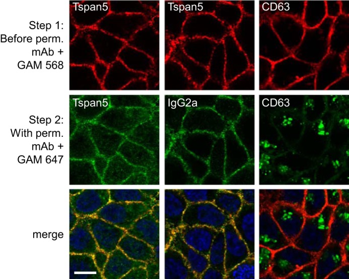
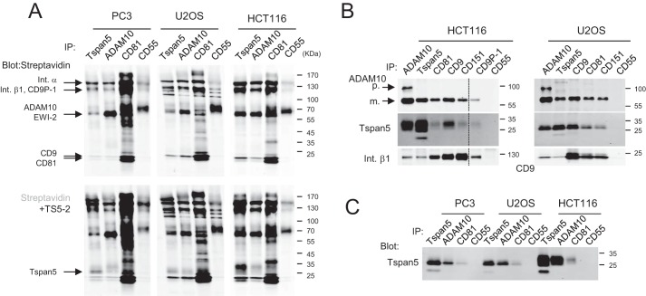
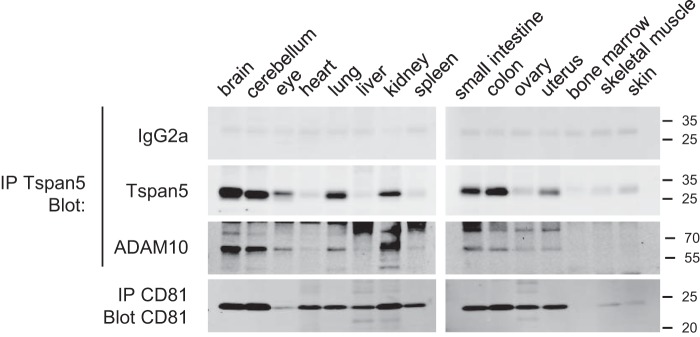
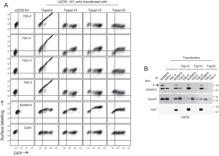
Tspan5 is a member of a subgroup of tetraspanins referred to as TspanC8. These tetraspanins directly interact with the metalloprotease ADAM10, regulate its exit from the endoplasmic reticulum and subsequent trafficking, and differentially regulate its ability to cleave various substrates and activate Notch signaling. The study of Tspan5 has been limited by the lack of good antibodies. This study provides new insights into Tspan5 using new monoclonal antibodies (mAbs), including two mAbs recognizing both Tspan5 and the highly similar tetraspanin Tspan17. Using these mAbs, we show that endogenous Tspan5 associates with ADAM10 in human cell lines and in mouse tissues where it is the most abundant, such as the brain, the lung, the kidney, or the intestine. We also uncover two TspanC8-specific motifs in the large extracellular domain of Tspan5 that are important for ADAM10 interaction and exit from the endoplasmic reticulum. One of the anti-Tspan5 mAbs does not recognize Tspan5 associated with ADAM10, providing a convenient way to measure the fraction of Tspan5 not associated with ADAM10. This fraction is minor in the cell lines tested, and it increases upon transfection of cells with TspanC8 tetraspanins such as Tspan15 or Tspan33 that inhibit Notch signaling. Finally, two antibodies inhibit ligand-induced Notch signaling, and this effect is stronger in cells depleted of the TspanC8 tetraspanin Tspan14, further indicating that Tspan5 and Tspan14 can compensate for each other in Notch signaling.

Keywords: ADAM; ADAM10; Notch pathway; Tspan5; intracellular trafficking; metalloprotease; monoclonal antibody; tetraspanin.


Conflict of interest statement

The authors declare that they have no conflicts of interest with the contents of this article

Figures

Figure 1.
Figure 1.
Characterization of new anti-Tspan5 mAb. A, flow cytometry analysis of U2OS cells expressing Tspan5 GFP and stained or not with mAbs to ADAM10, CD81, or three anti-Tspan5 mAbs. B, U2OS cells expressing Tspan5 GFP were lysed in RIPA or Brij 97 lysis buffer as indicated, before immunoprecipitation (IP) of Tspan5 using GFP trap beads. The samples were analyzed by Western blotting using the indicated antibodies. Note that the interaction of Tspan5 with ADAM10 is disrupted in RIPA buffer. C, binding of mAb TS5-2 to HCT116 was analyzed by flow cytometry 3 days after transfection with a control siRNA or two siRNA targeting Tspan5. D, HCT116 cells were lysed 3 days after transfection with a control siRNA or two Tspan5 siRNAs. In the top panels, the cells were lysed directly in Laemmli buffer, before Western blotting using a combination of anti-Tspan5 mAb TS5-2 and a secondary antibody. In the bottom panel, the cells were lysed in Brij 97 buffer, and Tspan5 was immunoprecipitated using TS5-2. The presence of Tspan5 in the immunoprecipitate was determined using a combination of biotin-labeled TS-2 mAb and streptavidin. E, mouse colon CT26 cells were lysed 3 days after transfection with a control siRNA or two to Tspan5 siRNAs. The cells were lysed directly in Laemmli buffer, before Western blotting using a combination of anti-Tspan5 mAb TS5-2 and a secondary antibody.
Figure 2.
Figure 2.
Specificity of selected anti-Tspan5 mAbs. A, U2OS cells stably expressing or not GFP-tagged Tspan5, Tspan14, Tspan15, and Tspan33 were lysed in RIPA buffer before immunoprecipitation (IP) of the transfected protein with GFP trap beads. The recognition of the transfected tetraspanin by the different mAbs was tested by Western blotting using both the cell lysate or the GFP-trap immunoprecipitate (IP). To control for the expression of the different transfected tetraspanins, the membrane was also incubated with an anti-GFP mAb. B, HeLa cells were transiently transfected with Tspan5, Tspan17, Tspan10, or CD9 and lysed 2 days later in Laemmli buffer. To control for the expression of the different transfected tetraspanins, the membrane was also incubated with an anti-GFP mAb. C, U2OS cells were transiently transfected with plasmids coding Tspan17 or Tspan5 and immunolabeled 2 days later with mAb TS5-2 and TS5/17. Note that the mAb TS5/17 also recognizes Tspan17 but TS5-2 does not (bar: 10 μm). D, U2OS cells were transiently transfected with plasmids coding the GFP-tagged chimeric tetraspanin Ts15LEL5, in which the LEL of Tspan15 was replaced by that of Tspan5 and the reverse chimera Ts5LEL15. The panel shows flow cytometry analysis of the binding of selected anti-Tspan5 mAb to the transfected cells.
Figure 3.
Figure 3.
Confocal microscopy analysis of the intracellular pool of Tspan5 and CD63. HCT116 cells were grown for 2 days on coverslips before incubation with anti-Tspan5 (TS5-2) or CD63 mAb for 1 h at 4 °C. After washing and fixation with paraformaldehyde, the cells were incubated with an anti-mouse polyclonal antibody coupled to Alexa 568 to visualize the surface pool of the tetraspanin (red). In Step 2, the cells were incubated with either the same primary antibody or a control IgG2a mAb in the presence of saponin to permeabilize the cells, and subsequently with an anti-mouse polyclonal antibody coupled to Alexa 647 (green). In this experiment, the surface pool of Tspan5 or CD63 is labeled with the two secondary antibodies, whereas the internal pool is labeled only with the Alexa 647-coupled secondary antibody (green). This experiment has been performed twice with similar outcomes. Perm., permeabilization; GAM, goat anti-mouse antibody. Bar, 10 μm.
Figure 4.
Figure 4.
Endogenous Tspan5 associates with ADAM10 and other tetraspanins. PC3, U2OS, and HCT116 were lysed in Brij 97, and immunoprecipitations (IP) were performed as indicated on the top of each lane. A, surface proteins were labeled with biotin before lysis and were visualized using Alexa 680-labeled streptavidin (top). In the 2nd step, the membrane was incubated with biotin-labeled TS5-2 mAb and again with Alexa 680-labeled streptavidin to confirm the presence of Tspan5 in the immunoprecipitates and compare its molecular weight with the other proteins present in the different immunoprecipitations (bottom). B, immunoprecipitations were performed from non-labeled cells. The composition of the immunoprecipitates was analyzed by Western blotting using various biotin-labeled mAbs. The mature (m.) and proform (p.) forms of ADAM10 are indicated by arrows. C, comparison of the migration profile of Tspan5 immunoprecipitated from PC3, U2OS, and HCT116 cells. Int., integrin.
Figure 5.
Figure 5.
Tspan5 associates with ADAM10 in mouse organs. Mouse organs were lysed, and immunoprecipitations (IP) were performed using Tspan5 or CD81 antibodies. The presence of the target antigen was analyzed by Western blotting using biotin-labeled mAbs. Because Tspan5 co-migrates with a nonspecific band, the membranes were incubated with an irrelevant biotin-labeled mAb of the same subclass (IgG2a) before incubation with the Tspan5 mAb. The presence of ADAM10 in the Tspan5 immunoprecipitates was analyzed using a polyclonal antibody to ADAM10. This experiment has been done twice with similar outcomes.
Figure 6.
Figure 6.
The majority of Tspan5 molecules associates with ADAM10. A, HCT116 cells were lysed in the presence of Brij 97, digitonin, and RIPA buffer before immunoprecipitations (IP) with the anti-ADAM10 mAb 11G2 or the anti-Tspan5 mAbs TS5-2 and TS5-1r. The presence of Tspan5 and ADAM10 in the immunoprecipitates was visualized by immunoblotting using biotin-labeled TS5-2 and 11G2 mAbs. Note that TS5-1r does not co-immunoprecipitate ADAM10 and that its ability to immunoprecipitate Tspan5 is poor under lysis conditions preserving the interaction of Tspan5 with ADAM10. The band corresponding to the proform (p.) of ADAM10 is indicated. B, U2OS-N1 cells stably expressing GFP-tagged Tspan5 were lysed in the presence of Brij 97 or digitonin before immunoprecipitations with the anti-ADAM10 mAb 11G2 or the anti-Tspan5 mAbs TS5-2 and TS5-1r. The presence of Tspan5 and ADAM10 in the immunoprecipitates was visualized by immunoblotting using biotin-labeled TS5-2 and 11G2 mAbs. Note that TS5-1r does not co-immunoprecipitate ADAM10. C, HCT116 and U2OS cells were transfected or not with a control siRNA or an siRNA targeting ADAM10 and lysed 3 days later in Brij 97. Immunoprecipitations were performed as indicated at the top of each lane. The presence of Tspan5 and ADAM10 in the immunoprecipitates was visualized by immunoblotting using biotin-labeled TS5-2 and 11G2 mAbs. Note the change of Tspan5 molecular weight upon ADAM10 silencing and the better immunoprecipitation of Tspan5 by TS5-1r. The band corresponding to the proform (p.) of ADAM10 is indicated. D, HCT116 or U2OS-N1 cells were lysed in digitonin and were subjected to two rounds of immunoprecipitation with a control mAb or the anti-ADAM10 mAb 11G2 or the anti-Tspan5 mAb TS5-2. Each sample was then separated in two for immunoprecipitations with the anti-ADAM10 and the anti-Tspan5 mAbs. The presence of Tspan5 and ADAM10 in the immunoprecipitates was visualized by immunoblotting using biotin-labeled TS5-2 and 11G2 mAbs. Except for B, the experiments in this figure have been done three times with similar outcomes.
Figure 7.
Figure 7.
ADAM10 facilitates Tspan5 exit from the ER. A, HCT116 and U2OS cells were transfected or not with a control siRNA or an siRNA targeting ADAM10 and grown for 3 days before lysis in Brij 97, and immunoprecipitations with mAb TS5-2 were performed. The immunoprecipitated proteins were treated or not with PNGase F or EndoH, as indicated before electrophoresis and immunoblotting using biotin-labeled TS5-2 mAb. Note that the lower molecular weight form of Tspan5 reinforced after ADAM10 silencing is EndoH-sensitive. B, binding of mAb TS5-2 and TS5-1r to HCT116 and U2OS cells, as well as that of the anti-ADAM10 mAb 11G2, was analyzed by flow cytometry 3 days after transfection with a control siRNA or an siRNA targeting ADAM10. Note the decrease of the binding of TS5-2 and the increase of the binding of TS5-1r. The dashed line corresponds to the labeling with only the secondary reagent. C, HCT116 cells treated or not with BFA were lysed using Brij 97 before immunoprecipitations with the anti-ADAM10 mAb 11G2 or the anti-Tspan5 mAb TS5-2 and TS5-1r. The presence of Tspan5 and ADAM10 in the immunoprecipitates was visualized by immunoblotting using biotin-labeled TS5-2 and 11G2 mAbs. Note that TS5-2 co-immunoprecipitates the proform of ADAM10 after BFA treatment and that ADAM10 co-immunoprecipitates the immature form of Tspan5. The experiments in this figure have been done twice.
Figure 8.
Figure 8.
Mutations of TspanC8-specific motifs in the LEL of Tspan5 abolish the interaction with ADAM10. A, sequence alignment of the LEL of CD151 and of the different Homo sapiens (Tspan), D. melanogaster (Tsp), and C. elegans (Tsp-12) TspanC8 tetraspanins. The residues present in most tetraspanins are in pink. The residues conserved in TspanC8 are also indicated: red, >80% conservation; blue, >60% conservation; green, conservative substitutions. The two additional cysteines that are the hallmark of TspanC8 are in yellow. B, HeLa cells were transfected with GFP-tagged Tspan5 or the different mutants as indicated at the top of each lane. Two days later, the cells were lysed in Brij 97, and immunoprecipitations were performed as indicated. The presence of ADAM10, Tspan5, or the mutants in the immunoprecipitates was analyzed by Western blotting. The mature (m.) form and the proform (p.) of ADAM10 are indicated. C, HeLa cells were transfected with plasmids encoding GFP-tagged Tspan5 or the RDD and DID mutants. Two days later, the surface expression of ADAM10 and the binding of the anti-Tspan5 mAb TS5-2 to the cells were analyzed by flow cytometry. The experiments in this figure have been done twice.
Figure 9.
Figure 9.
Two Tspan5 mutants that do not interact with ADAM10 are retained in the endoplasmic reticulum. A, HeLa cells were co-transfected with plasmids encoding the indicated GFP-tagged Tspan5 or Tspan15 mutants and a plasmid encoding mCherry-tagged Sec61, an ER marker. The images were acquired by confocal microscopy. B, immunofluorescence microscopy analysis of HeLa cells transfected with GFP-tagged Tspan5 mutants RDD and NIYF and stained with mAbs TS5-2 and TS5/17 after Triton X-100 permeabilization. Bar: 10 μm.
Figure 10.
Figure 10.
Effect of Tspan5 mAbs on ligand-induced Notch signaling. A, Notch activity in U2OS-N1, measured using a luciferase reporter assay. U2OS-N1 cells treated or not with the indicated mAbs were co-cultured with OP9-DLL1 cells for 20–24 h to activate Notch signaling or with parental OP9 cells to determine the basal level of luciferase production. The graph shows the mean ± S.E. of 3–7 independent experiments in duplicate. The data are expressed as a percentage of the signal observed for control U2OS-N1 cells. B, Notch activity, measured using a luciferase reporter assay, of U2OS-N1 cells treated with the indicated siRNAs. Notch was activated by incubation with OP9-DLL1 cells. The graph shows the mean ± S.E. of six independent experiments performed in duplicate. C, Notch activity, measured using a luciferase reporter assay, of U2OS-N1 cells treated with a control siRNA or an siRNA to Tspan14, and the indicated mAbs. The graph shows the mean ± S.E. of 3–6 independent experiments performed in duplicate. ***, p < 0.001; **, p < 0.01; *, p < 0.05 as compared with control.
Figure 11.
Figure 11.
Ectopically expressed TspanC8 tetraspanins compete with endogenous Tspan5 for the association with ADAM10. A, flow cytometry analysis of U2OS cells stably expressing GFP-tagged Tspan5, Tspan14, Tspan15, or Tspan33 and stained or not with mAbs to ADAM10, CD81, or four anti-Tspan5 mAb. Note the increase in TS5-1r binding proportionally to the GFP signal and the slight decrease of the binding of TS5-2 and TS5-3. B, U2OS cells stably expressing or not GFP-tagged Tspan14, Tspan15, or Tspan33 were lysed in digitonin before immunoprecipitations (IP) with the anti-ADAM10 mAb 11G2 or the anti-Tspan5 mAbs TS5-2 and TS5-1r. The presence of Tspan5 and ADAM10 in the immunoprecipitates was visualized by immunoblotting using biotin-labeled TS5-2 and 11G2 mAbs. The band corresponding to the proform (p.) of ADAM10 is indicated. All experiments in this figure have been done three times with similar outcomes.

Similar articles

Cited by

References

    1. Charrin S., Jouannet S., Boucheix C., and Rubinstein E. (2014) Tetraspanins at a glance. J. Cell Sci. 127, 3641–3648 - PubMed
    1. Charrin S., le Naour F., Silvie O., Milhiet P. E., Boucheix C., and Rubinstein E. (2009) Lateral organization of membrane proteins: tetraspanins spin their web. Biochem. J. 420, 133–154 - PubMed
    1. Hemler M. E. (2005) Tetraspanin functions and associated microdomains. Nat. Rev. Mol. Cell Biol. 6, 801–811 - PubMed
    1. Yáñez-Mó M., Barreiro O., Gordon-Alonso M., Sala-Valdés M., and Sánchez-Madrid F. (2009) Tetraspanin-enriched microdomains: a functional unit in cell plasma membranes. Trends Cell Biol. 19, 434–446 - PubMed
    1. Serru V., Dessen P., Boucheix C., and Rubinstein E. (2000) Sequence and expression of seven new tetraspans. Biochim. Biophys. Acta 1478, 159–163 - PubMed

Publication types

MeSH terms

LinkOut - more resources