. 2017 Jun 9;292(23):9599-9612.
doi: 10.1074/jbc.M116.746263. Epub 2017 Apr 20.

Crystal structure and redox properties of a novel cyanobacterial heme protein with a His/Cys heme axial ligation and a Per-Arnt-Sim (PAS)-like domain

Affiliations

Crystal structure and redox properties of a novel cyanobacterial heme protein with a His/Cys heme axial ligation and a Per-Arnt-Sim (PAS)-like domain

Taiki Motomura et al. J Biol Chem. .

Abstract

Photosystem II catalyzes light-induced water oxidation leading to the generation of dioxygen indispensable for sustaining aerobic life on Earth. The Photosystem II reaction center is composed of D1 and D2 proteins encoded by psbA and psbD genes, respectively. In cyanobacteria, different psbA genes are present in the genome. The thermophilic cyanobacterium Thermosynechococcus elongatus contains three psbA genes: psbA1, psbA2, and psbA3, and a new c-type heme protein, Tll0287, was found to be expressed in a strain expressing the psbA2 gene only, but the structure and function of Tll0287 are unknown. Here we solved the crystal structure of Tll0287 at a 2.0 Å resolution. The overall structure of Tll0287 was found to be similar to some kinases and sensor proteins with a Per-Arnt-Sim-like domain rather than to other c-type cytochromes. The fifth and sixth axial ligands for the heme were Cys and His, instead of the His/Met or His/His ligand pairs observed for most of the c-type hemes. The redox potential, E½, of Tll0287 was -255 ± 20 mV versus normal hydrogen electrode at pH values above 7.5. Below this pH value, the E½ increased by ≈57 mV/pH unit at 15 °C, suggesting the involvement of a protonatable group with a pKred = 7.2 ± 0.3. Possible functions of Tll0287 as a redox sensor under microaerobic conditions or a cytochrome subunit of an H2S-oxidizing system are discussed in view of the environmental conditions in which psbA2 is expressed, as well as phylogenetic analysis, structural, and sequence homologies.

Keywords: D1 protein; His-Cys heme axial coordination; PAS domain; PAS-like domain; Tll0287; X-ray crystallography; cytochrome; heme; photosynthesis; photosystem II.


Conflict of interest statement

The authors declare that they have no conflicts of interest with the contents of this article

Figures

Figure 1.
Figure 1.
Immunoblot analysis on the expression and location of Tll0287. a, SDS-PAGE analysis with a 16% acrylamide gel of the purified Tll0287 protein (lane 2) used in this study. The molecular mass markers are shown in lane 1, with their molecular masses (kDa) indicated. b, CBB-stained gel of the samples used for immunoblot analysis shown in c. The samples shown are the purified Tll0287 protein (Tll), whole cell, and thylakoid fractions from the WT (predominately expressing psbA1), WT*2 (expressing psbA2 only), and WT*3 (expressing psbA3 only) strains, and crude-PSII (LDAO-PSII), PSII core complex purified from the psbA2-expressing strain (WT*2). The amount of proteins loaded for each lane is equivalent to 2 μg of chlorophyll for the whole cell fractions and thylakoid membranes, 1 μg of chlorophyll for LDAO-PSII, 0.5 μg for purified PSII, and 0.16 mg of protein for the Tll0287 protein. M stands for molecular marker with the molecular masses indicated (kDa). c, immunoblot analysis with an anti-Tll0287 antibody for same samples shown in b.
Figure 2.
Figure 2.
Digestion of the Tll0287 protein with a protease thermolysin. a, CBB-stained gel of the samples used for immunoblot analysis shown in b and c. The samples used are the purified Tll0287 protein, thylakoid membrane fraction from the WT*2 strain (A2-Thy, expressing psbA2 only), and purified PSII from WT*2 (A2-PSII). The amount of the samples loaded on each lane was the same as that in Fig. 1b. b, immunoblot analysis of the Tll0287 protein in the purified Tll0287 sample and thylakoid membrane fraction (A2-Thy) from the WT*2 strain with an anti-Tll0287 antibody, either before (labeled −) or after digestion of the samples with 5 μg/ml thermolysin for 6 h (labeled +TH). c, immunoblot analysis of the PsbO protein in the purified PSII and thylakoid membrane fraction from the WT*2 strain with an anti-PsbO antibody, before (−) or after digestion of the samples with thermolysin (+TH) at the same conditions as for Tll0287.
Figure 3.
Figure 3.
Electrochemistry on Tll0287. a and b show the absorption spectra recorded at the indicated potential during the oxidative and reductive processes, respectively. c shows a Nernst plot for an oxidation (blue points) and a reduction (red points) performed at pH 8.5 and T = 15 °C. The continuous black line is a global fit with n fixed to 1 and E½′ = −255 mV versus NHE. d shows the pH dependence of the E½′ determined as above at different pH values. The continuous line is the result of a fitting procedure with the following equation: E½′ = E0′ − 57 log((Kox + [H+])/(Kred + [H+])), where E0′ is the midpoint redox potential of the fully protonated form, and Kred and Kox are the ionization constants of the protonatable group when the iron is reduced and oxidized, respectively. The pKred was found to be 7.2.
Figure 4.
Figure 4.
Crystal structure of the Tll0287 protein. a, overall structure of the Tll0287 protein. The PAS-like domain composed of three α-helices and four β-strands is depicted in green; the heme and its binding part are depicted in red and yellow, respectively; and the loop region is depicted in orange. b, surface charge distribution of the Tll0287 protein. Blue and red represent negative and positive areas, respectively. The area surrounding the heme is largely positive. The black arrow indicates a concave pocket. c, ligand environment of the heme c in Tll0287. The fifth and sixth heme iron axial ligands, Cys68 and His145, and the residues around them are depicted in stick models. d, hydrophobicity characters of the surface of Tll0287. The structure of Tll0287 was depicted in a ball model in the same direction (left side) as that in panels a and b, and in a direction rotated 180° (right side). Blue represents positively charged residues (Arg, Lys, and His), red represents negatively charged residues (Asp and Glu), white represents hydrophilic residues, and yellow represents hydrophobic residues.
Figure 5.
Figure 5.
Expression level of Tll0287 under dark conditions. Spectrum a, WT whole cells cultivated for 2 days under normal light conditions. Spectrum b, WT whole cells after a further dark period of 3–4 h. Spectrum c, WT*2 whole cells cultivated for 2 days under normal light conditions. Spectrum d, WT*2 whole cells after a further dark period of 3–4 h. For sake of comparison, the spectra were approximately scaled by using both the Fe3+SOD signals (labeled with asterisk between 1300 and 2000 gausses) and the gz signal of cyt c-550 (labeled with asterisk between 2200 and 2300 gausses). The signal from Tll0287 is located at ∼2800 gauss. Instrument settings were as follows: modulation amplitude, 25 G; microwave power, 5 milliwatts; temperature, 20 K; microwave frequency, 9.4 GHz; and modulation frequency, 100 kHz.
Figure 6.
Figure 6.
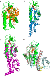
Structural comparison of the Tll0287 protein with other proteins. a, superposition of Tll0287 and PsbV2 from T. elongatus (PDB code 4LJI) (37). The colors for the Tll0287 protein and its heme are green and red, respectively, and those for the PsbV2 protein are as follows: heme, yellow; others, orange. b, superposition of Tll0287 and the sporulation kinase D sensor domain from B. subtilis (blue, A chain of PDB code 4JGO; pink, the ligand of this protein, pyruvate) (48). c, superimposition of Tll0287 and a methyl-accepting chemotaxis protein (Mcp) (purple, A chain of PDB code 3C8C; cyan, the ligand of this protein, alanine). d, superimposition of Tll0287 and the dimer of the non-membrane part of GSU0935 (PDB code 3B42) (62).
Figure 7.
Figure 7.
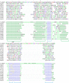
Structural alignment of the Tll0287 protein with 11 homologous proteins. Eleven representative proteins are selected from supplemental Table S1 that show structural similarities with Tll0287, excluding their similar molecules. Inserted segments relative to the Tll0287 protein structure are hidden. The upper part shows the alignment of the amino acid sequence, and the lower part shows the secondary structure assignments performed by DSSP (65, 66). H, helix; E, strand; L, coil.
Figure 8.
Figure 8.
A possible extra ligand in Tll0287. a, the residues Arg87, Asp105, and Thr179 surrounding the concave pocket (shown in Fig. 4b) are depicted as stick models. b, electron density map of a possible ligand corresponding to the ligand binding site in the structure of the sporulation kinase D sensor domain (48). The blue mesh and red mesh represent mFo − DFc omit map (positive and negative, respectively) contoured at 3.5 σ distributions, and the gray mesh represents 2mFo − DFc map contoured at 1.0 σ distributions.

Similar articles

Cited by

References

    1. Umena Y., Kawakami K., Shen J.-R., and Kamiya N. (2011) Crystal structure of oxygen-evolving photosystem II at a resolution of 1.9 Å. Nature 473, 55–60 - PubMed
    1. Suga M., Akita F., Hirata K., Ueno G., Murakami H., Nakajima Y., Shimizu T., Yamashita K., Yamamoto M., Ago H., and Shen J.-R. (2015) Native structure of photosystem II at 1.95 Å resolution viewed by femtosecond X-ray pulses. Nature 517, 99–103 - PubMed
    1. Wydrzynski T. J., and Satoh K. (eds.) (2005) Photosystem II, the Light-Driven Water: Plastoquinone Oxidoreductase, Springer, Dordrecht, The Netherlands
    1. Shen J.-R. (2015) Structure of Photosystem II and the mechanism of water oxidation in photosynthesis. Annu. Rev. Plant Biol. 66, 23–48 - PubMed
    1. Kawakami K., Umena Y., Kamiya N., and Shen J.-R. (2011) Structure of the catalytic, inorganic core of oxygen-evolving Photosystem II at 1.9 Å resolution. J. Photochem. Photobiol. B 104, 9–18 - PubMed

MeSH terms

LinkOut - more resources