

Epithelial-to-Mesenchymal Transition Contributes to Immunosuppression in Breast Carcinomas
- PMID: 28428275
- PMCID: PMC5541771
- DOI: 10.1158/0008-5472.CAN-16-3292
Epithelial-to-Mesenchymal Transition Contributes to Immunosuppression in Breast Carcinomas
Abstract
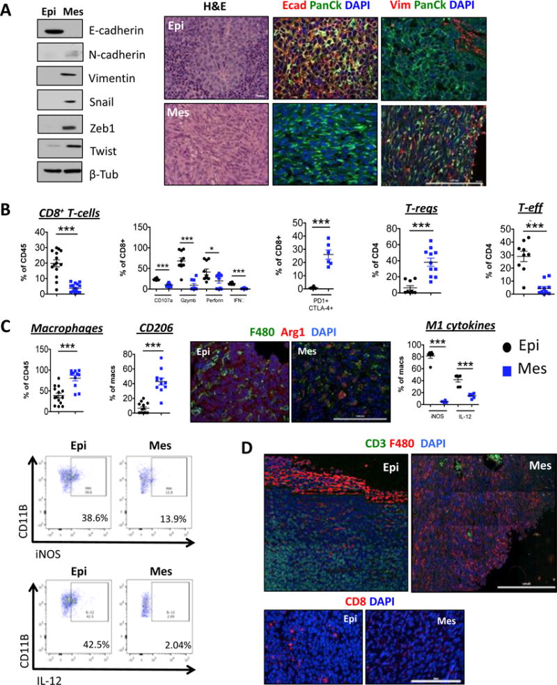
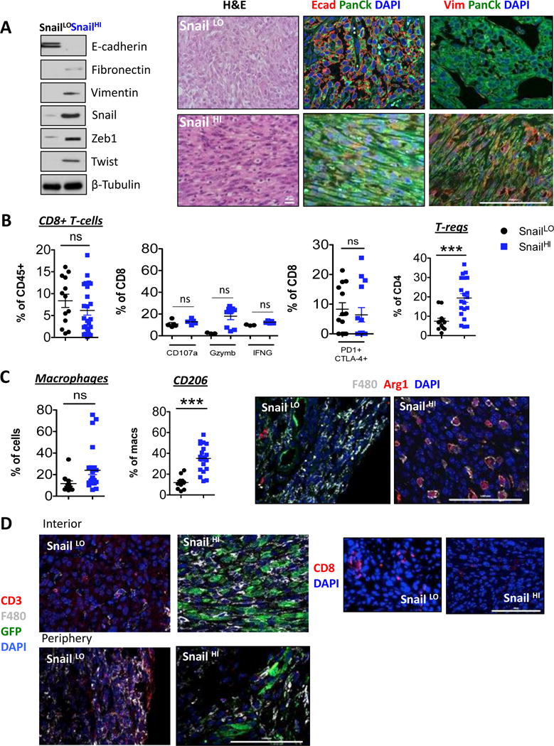
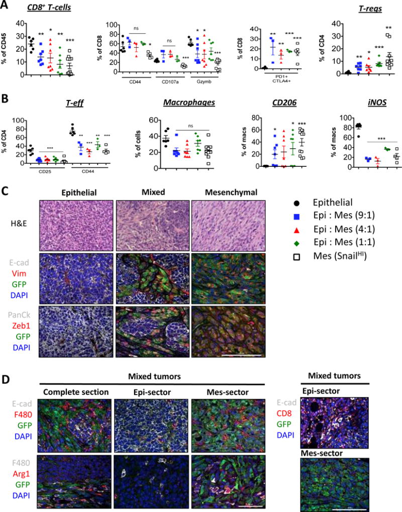
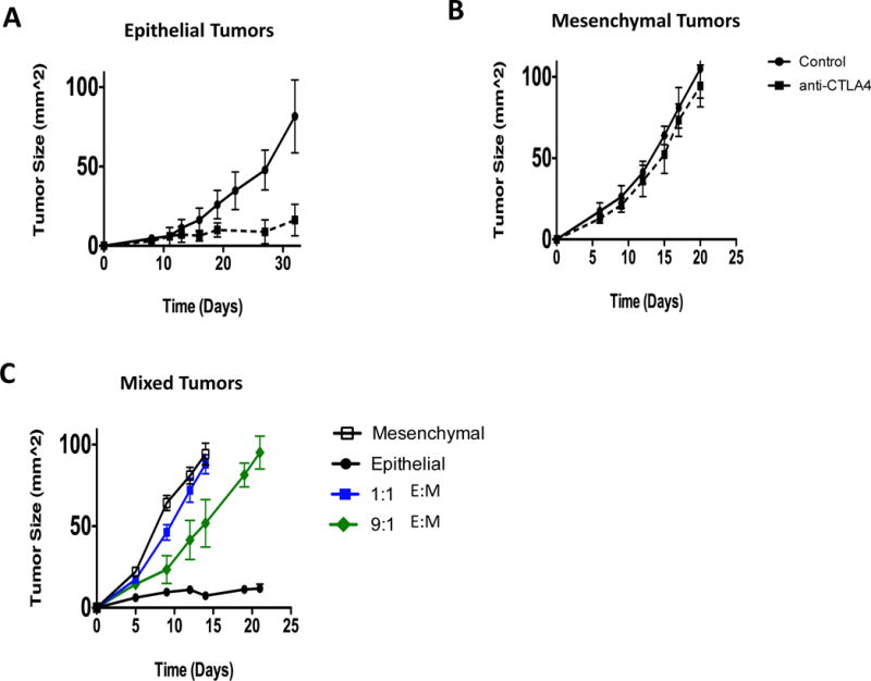
The epithelial-to-mesenchymal transition (EMT) is a cell biological program that confers mesenchymal traits on carcinoma cells and drives their metastatic dissemination. It is unclear, however, whether the activation of EMT in carcinoma cells can change their susceptibility to immune attack. We demonstrate here that mammary tumor cells arising from more epithelial carcinoma cell lines expressed high levels of MHC-I, low levels of PD-L1, and contained within their stroma CD8+ T cells and M1 (antitumor) macrophages. In contrast, tumors arising from more mesenchymal carcinoma cell lines exhibiting EMT markers expressed low levels of MHC-I, high levels of PD-L1, and contained within their stroma regulatory T cells, M2 (protumor) macrophages, and exhausted CD8+ T cells. Moreover, the more mesenchymal carcinoma cells within a tumor retained the ability to protect their more epithelial counterparts from immune attack. Finally, epithelial tumors were more susceptible to elimination by immunotherapy than corresponding mesenchymal tumors. Our results identify immune cells and immunomodulatory markers that can be potentially targeted to enhance the susceptibility of immunosuppressive tumors to various therapeutic regimens. Cancer Res; 77(15); 3982-9. ©2017 AACR.
©2017 American Association for Cancer Research.
Figures
Similar articles
-
Epithelial-to-mesenchymal transition and autophagy induction in breast carcinoma promote escape from T-cell-mediated lysis.Cancer Res. 2013 Apr 15;73(8):2418-27. doi: 10.1158/0008-5472.CAN-12-2432. Epub 2013 Feb 22. Cancer Res. 2013. PMID: 23436798
-
Epithelial-to-mesenchymal transition (EMT) induced by inflammatory priming elicits mesenchymal stromal cell-like immune-modulatory properties in cancer cells.Br J Cancer. 2015 Mar 17;112(6):1067-75. doi: 10.1038/bjc.2015.29. Br J Cancer. 2015. PMID: 25668006 Free PMC article.
-
Core needle biopsy of breast cancer tumors increases distant metastases in a mouse model.Neoplasia. 2014 Nov 20;16(11):950-60. doi: 10.1016/j.neo.2014.09.004. eCollection 2014 Nov. Neoplasia. 2014. PMID: 25425969 Free PMC article.
-
Immunological Consequences of Epithelial-Mesenchymal Transition in Tumor Progression.J Immunol. 2016 Aug 1;197(3):691-8. doi: 10.4049/jimmunol.1600458. J Immunol. 2016. PMID: 27431984 Free PMC article. Review.
-
Immunosuppressive cells in tumor immune escape and metastasis.J Mol Med (Berl). 2016 May;94(5):509-22. doi: 10.1007/s00109-015-1376-x. Epub 2015 Dec 22. J Mol Med (Berl). 2016. PMID: 26689709 Review.
Cited by
-
Plasticity-induced repression of Irf6 underlies acquired resistance to cancer immunotherapy in pancreatic ductal adenocarcinoma.Nat Commun. 2024 Feb 20;15(1):1532. doi: 10.1038/s41467-024-46048-7. Nat Commun. 2024. PMID: 38378697 Free PMC article.
-
EMT-Associated Heterogeneity in Circulating Tumor Cells: Sticky Friends on the Road to Metastasis.Cancers (Basel). 2020 Jun 19;12(6):1632. doi: 10.3390/cancers12061632. Cancers (Basel). 2020. PMID: 32575608 Free PMC article. Review.
-
Mesenchymal ovarian cancer cells promote CD8+ T cell exhaustion through the LGALS3-LAG3 axis.NPJ Syst Biol Appl. 2023 Dec 12;9(1):61. doi: 10.1038/s41540-023-00322-4. NPJ Syst Biol Appl. 2023. PMID: 38086828 Free PMC article.
-
Concomitant attenuation of HMGCR expression and activity enhances the growth inhibitory effect of atorvastatin on TGF-β-treated epithelial cancer cells.Sci Rep. 2021 Jun 17;11(1):12763. doi: 10.1038/s41598-021-91928-3. Sci Rep. 2021. PMID: 34140545 Free PMC article.
-
E-liquid alters oral epithelial cell function to promote epithelial to mesenchymal transition and invasiveness in preclinical oral squamous cell carcinoma.Sci Rep. 2023 Feb 27;13(1):3330. doi: 10.1038/s41598-023-30016-0. Sci Rep. 2023. PMID: 36849550 Free PMC article.
References
-
- Hanahan D, Coussens LM. Accessories to the crime: functions of cells recruited to the tumor microenvironment. Cancer Cell. 2012;21(3):309–22. - PubMed
-
- Viel S, Marcais A, Guimaraes FS, Loftus R, Rabilloud J, Grau M, et al. TGF-beta inhibits the activation and functions of NK cells by repressing the mTOR pathway. Sci Signal. 2016;9(415):ra19. - PubMed
Publication types
MeSH terms
Grants and funding
LinkOut - more resources
Full Text Sources
Other Literature Sources
Medical
Research Materials
