. 2017 Apr 20;7(1):989.
doi: 10.1038/s41598-017-01022-w.

Poly-protein G-expressing bacteria enhance the sensitivity of immunoassays

Affiliations

Poly-protein G-expressing bacteria enhance the sensitivity of immunoassays

Wen-Rui Hao et al. Sci Rep. .

Erratum in

Abstract

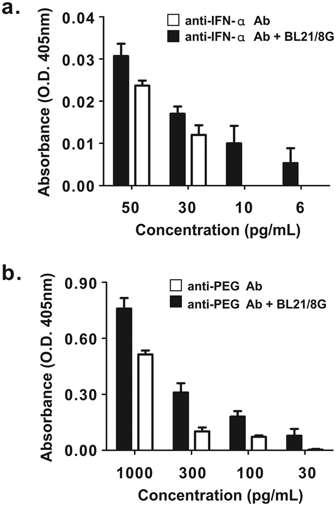
The sensitivities of solid-phase immunoassays are limited by the quantity of detection antibodies bound to their antigens on the solid phase. Here, we developed a poly-protein G-expressing bacterium as an antibody-trapping microparticle to enhance the signals of immunoassays by increasing the accumulation of detection antibodies on the given antigen. Eight tandemly repeated fragment crystallisable (Fc) binding domains of protein G were stably expressed on the surface of Escherichia coli BL21 cells (termed BL21/8G). BL21/8G cells showed a higher avidity for trapping antibodies on their surface than monomeric protein G-expressing BL21 (BL21/1G) cells did. In the sandwich enzyme-linked immunosorbent assay (ELISA), simply mixing the detection antibody with BL21/8G provided a detection limit of 6 pg/mL for human interferon-α (IFN-α) and a limit of 30 pg/mL for polyethylene glycol (PEG)-conjugated IFN-α (Pegasys), which are better than that of the traditional ELISA (30 pg/mL for IFN-α and 100 pg/mL for Pegasys). Moreover, the sensitivity of the Western blot for low-abundance Pegasys (0.4 ng/well) was increased by 25 folds upon mixing of an anti-PEG antibody with BL21/8G cells. By simply being mixed with a detection antibody, the poly-protein G-expressing bacteria can provide a new method to sensitively detect low-abundance target molecules in solid-phase immunoassays.


Conflict of interest statement

The authors declare that they have no competing interests.

Figures

Figure 1
Figure 1
Poly-protein G-expressing BL21 bacteria. (a) Strategy for the non-covalent modification of detection antibodies to the single protein G C2 domain-expressing bacteria (BL21/1G) or the eight tandemly repeated C2 domain-expressing bacteria (BL21/8G). (b) Schematic representation of the gene construction of the protein G-expressing plasmids (1G and 8G). The construction includes, from N to C termini, an HA epitope tag, a single or eight tandemly repeated protein G C2 domain fragment (C2), a (GGGSG)3 linker (L), and an AIDA.
Figure 2
Figure 2
Expression of poly-protein G in BL21 bacteria. (a) The expression of poly-protein G in BL21 bacteria was analyzed by Western blot using a mouse anti-HA tag antibody. Lane 1, BL21 as negative control; Lane 2, single protein C2 domain-expressing BL21 bacteria (BL21/1G); Lane 3, eight tandemly repeated C2 domain-expressing BL21 bacteria (BL21/8G). (b) The transformed BL21 bacteria were induced by IPTG, and the growth rate of the bacteria was measured by recording the absorbance at 600 nm every two hours. (●)BL21/1G, (■)BL21/8G, (○)BL21/1G + IPTG, (□)BL21/8G + IPTG. Bar, SD.
Figure 3
Figure 3
Fluorescent microscopy of poly-protein G-expressing bacteria labeled with FITC-conjugated antibody. The antibody-trapping ability of BL21, BL21/1G and BL21/8G was analyzed by FITC-conjugated goat antibody under a fluorescent microscope. Top panels, green fluorescence of FITC under dark field. Bottom panels, images of bacteria under bright field. Bar = 10 μm.
Figure 4
Figure 4
The antibody-trapping ability of poly-protein G-expressing bacteria. (a) Equivalent amounts of BL21, BL21/1G and BL21/8G cells were coated on 96-well plates. The absorbance of the bacteria was measured at 600 nm. (b) The antibody-trapping ability of BL21, BL21/1G and BL21/8G cells coated on 96-well plates was analyzed by HRP-conjugated goat antibody. Statistical analysis was performed by one-way ANOVA. (c) BL21/1G and BL21/8G cells were preserved in PBS containing 30% (v/v) glycerol at indicated temperatures for 40 days. The antibody-trapping ability of these bacteria was analyzed by HRP-conjugated goat antibody. Statistical analysis was performed by two-way ANOVA. Bar, SD. ns, no significant difference. *P value < 0.05; **P value < 0.001.
Figure 5
Figure 5
ELISA tests performed in the presence and absence of poly-protein G-expressing bacteria. Anti-PEG antibody (6.3-biotin) mixed with BL21/1G cells or BL21/8G cells, or not mixed with anything, was added into 96-well plates coated with (a) PEG5k-BSA or (b) BSA. The binding amount of 6.3-biotin was determined by measuring absorbance at 405 nm after staining with SA-HRP and ABTS. Ab, 6.3-biotin. Statistical analysis was performed by one-way ANOVA. Bar, SD. ns, no significant difference. **P value < 0.001.
Figure 6
Figure 6
Detection of IFN-α and Pegasys by traditional sandwich ELISA or BL21/8G-based sandwich ELISA. (a) Serial diluted IFN-α samples were added into 96-well plates coated with anti-IFN-α capture antibody (MT1, IgM). Captured IFN-α molecules were detected by biotin-conjugated anti-IFN-α detection antibody (MT2, IgG) mixed with BL21/8G cells or not mixed with BL21/8G cells, and then color was developed using ABTS. (b) Serially diluted Pegasys samples were added into 96-well plates coated with anti-PEG capture antibody (AGP4, IgM). Captured Pegasys molecules were detected by biotin-conjugated anti-PEG detection antibody (3.3-biotin, IgG) mixed with BL21/8G cells or not mixed with BL21/8G cells, and color was developed using ABTS. Statistical analysis was performed by an independent t-test. Bar, SD.
Figure 7
Figure 7
Western blot of PEG-conjugated IFN-α (Pegasys). Serially diluted Pegasys samples were electrophoresed on a 10% (w/v) reducing SDS-PAGE, transferred to nitrocellulose membrane, and probed with anti-PEG antibody (3.3) mixed with BL21/8G cells (lower panel) or not mixed with BL21/8G cells (upper panel) as described in the Materials and Methods section.

References

    1. Bienvenut WV, et al. Toward a clinical molecular scanner for proteome research: Parallel protein chemical processing before and during Western blot. Anal. Chem. 1999;71:4800–4807. doi: 10.1021/ac990448m. - DOI - PubMed
    1. Kragstrup TW, Vorup-Jensen T, Deleuran B, Hvid M. A simple set of validation steps identifies and removes false results in a sandwich enzyme-linked immunosorbent assay caused by anti-animal IgG antibodies in plasma from arthritis patients. Springerplus. 2013;2:263–270. doi: 10.1186/2193-1801-2-263. - DOI - PMC - PubMed
    1. Frenzel C, et al. Evaluation of F-actin ELISA for the diagnosis of autoimmune hepatitis. Am. J. Gastroenterol. 2006;101:2731–2736. doi: 10.1111/j.1572-0241.2006.00830.x. - DOI - PubMed
    1. Fan P, et al. Enhanced sensitivity for detection of HIV-1 p24 antigen by a novel nuclease-linked fluorescence oligonucleotide assay. PLoS ONE. 2015;10:e0125701. doi: 10.1371/journal.pone.0125701. - DOI - PMC - PubMed
    1. Pesce AJ, Michael JG. Artifacts and limitations of enzyme immunoassay. J. Immunol. Methods. 1992;150:111–119. doi: 10.1016/0022-1759(92)90070-A. - DOI - PubMed

Publication types

MeSH terms