. 2017 Apr 20;7(1):974.
doi: 10.1038/s41598-017-01129-0.

The tarantula toxin β/δ-TRTX-Pre1a highlights the importance of the S1-S2 voltage-sensor region for sodium channel subtype selectivity

Affiliations

The tarantula toxin β/δ-TRTX-Pre1a highlights the importance of the S1-S2 voltage-sensor region for sodium channel subtype selectivity

Joshua S Wingerd et al. Sci Rep. .

Abstract

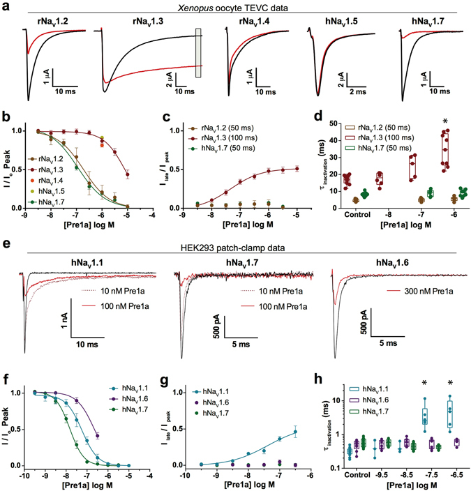
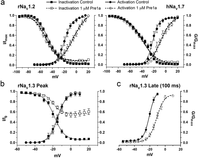
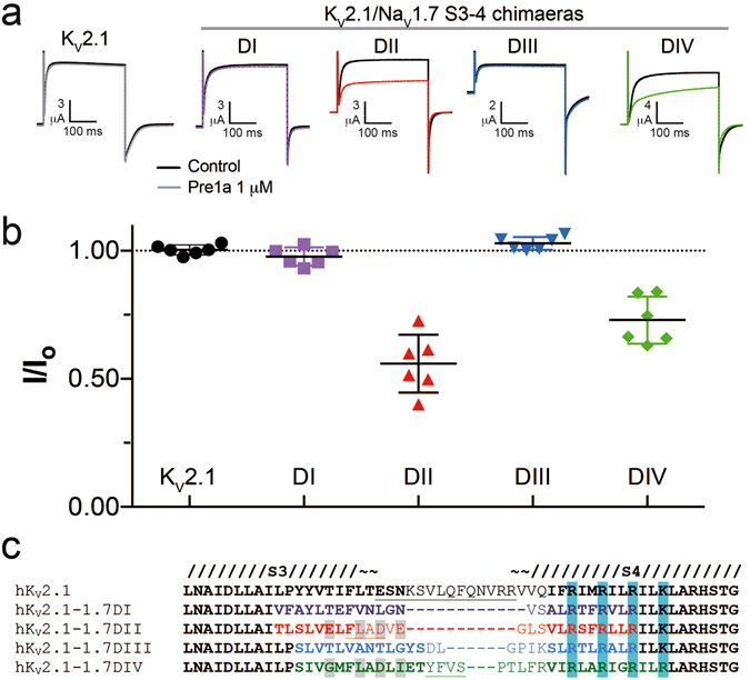
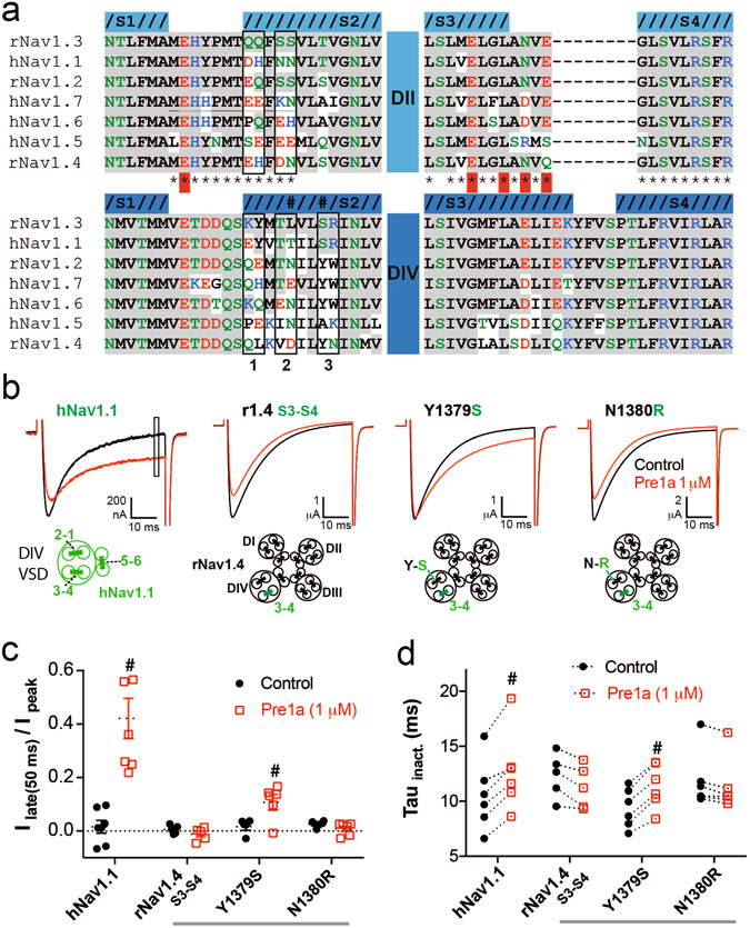
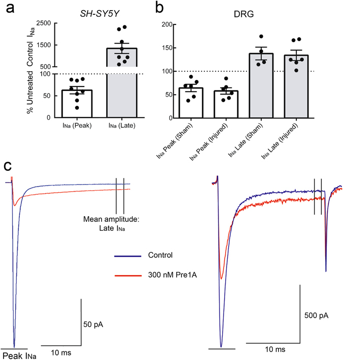
Voltage-gated sodium (NaV) channels are essential for the transmission of pain signals in humans making them prime targets for the development of new analgesics. Spider venoms are a rich source of peptide modulators useful to study ion channel structure and function. Here we describe β/δ-TRTX-Pre1a, a 35-residue tarantula peptide that selectively interacts with neuronal NaV channels inhibiting peak current of hNaV1.1, rNaV1.2, hNaV1.6, and hNaV1.7 while concurrently inhibiting fast inactivation of hNaV1.1 and rNaV1.3. The DII and DIV S3-S4 loops of NaV channel voltage sensors are important for the interaction of Pre1a with NaV channels but cannot account for its unique subtype selectivity. Through analysis of the binding regions we ascertained that the variability of the S1-S2 loops between NaV channels contributes substantially to the selectivity profile observed for Pre1a, particularly with regards to fast inactivation. A serine residue on the DIV S2 helix was found to be sufficient to explain Pre1a's potent and selective inhibitory effect on the fast inactivation process of NaV1.1 and 1.3. This work highlights that interactions with both S1-S2 and S3-S4 of NaV channels may be necessary for functional modulation, and that targeting the diverse S1-S2 region within voltage-sensing domains provides an avenue to develop subtype selective tools.


Conflict of interest statement

The authors declare that they have no competing interests.

Figures

Figure 1
Figure 1
Identification and sequence of TRTX-Pre1a. (a) RP-HPLC chromatogram of crude venom from P. reduncus indicating the fraction (F18) responsible for robust inhibition of hNaV1.7 expressed in Xenopus oocytes (inset). (b) Final analytical RP-HPLC purification step of F18 (TRTX-Pre1a) (inset: MALDI-TOF MS spectrum showing M + H+ of 4227.5). (c) Activity of pure, native Pre1a on rNaV1.3 and hNaV1.7 exressed in Xenopus oocytes demonstrating inhibition of inactivation and peak current, respectively. (d) Sequence alignment of TRTX-Pre1a with NaV modulating Theraphotoxins, , , , –, , –. Percent similarity was calculated comparing the number of identical (dark gray) and similar (light gray) amino acids.
Figure 2
Figure 2
Pre1a shows conformational flexibility under RP-HPLC conditions. Analytical RP-HPLC of pure synthetic Pre1a shows the presence of multiple conformers in and acetonitrile/water mixture at room temperature. Insets 1 and 2, demonstrate identical elution profiles for reinjection of two fractions (highlighted and numbered) taken from the major peak, discounting the presence of impurities.
Figure 3
Figure 3
Pre1a shows conformational flexibility in aqueous conditions. (a) 2D 1H-15N-HSQC of recombinantly produced Pre1a. The chemical shifts of resonances for residues in loop 1 (D2–R9, underlined) show multiple peaks indicating the presence of three conformations of the peptide (A = Major, B = middle, C = minor, highlighted for G5 in the inset). sc = side chain NH resonances for N, R and W residues. (b) Two views of a homology model of Pre1a (based on the NMR structure of HwTxIV, PDB: 2M4X) illustrating the relative positions of W6 and F7 at the tip of Loop 1 and W29, Y32 and Y21. Loop 1 residues (that show multiple peaks in the HSQC above) are in red, disulfide bonds are in yellow. The right panel highlights the position of W6 and F7 at the tip of Loop 1 and the positions of C3 and R9, which may act as a hinge region for movement of the loop.
Figure 4
Figure 4
Pre1a preferentially inhibits neuronal NaV channels. (a) Representative NaV currents recorded from Xenopus oocytes (α-subunit alone) before addition of 1 µM Pre1a (black) and after reaching steady state inhibition (red). Late current was assessed at 50 ms (100 ms for NaV1.3) from the peak current, as highlighted by the grey box on rNaV1.3. (b) Concentration-effect curves for peak current inhibition by Pre1a for rNaV1.2 (IC50 189.6 nM; n ≥ 5), rNaV1.3 (IC50 8.0 μM; n ≥ 8) and hNaV1.7 (IC50 114.0 nM; n ≥ 8), with single point 1 µM concentrations for rNaV1.4 (n = 6) and hNaV1.5 (n = 7). (c) Concentration-response curve for late current inhibition of inactivation by Pre1a of rNaV1.3 (EC50 45.0 nM). (d) Concentration-dependent effects of Pre1a on the rate of inactivation (τ) for rNaV1.3, with 1 µM demonstrating a significant slowing of inactivation (p < 0.001; n ≥ 5; ANOVA with Dunnet’s test). (e) Representative current traces of hNaV1.1, hNaV1.6, and hNaV1.7 expressed in HEK cells co-expressed with NaVβ1, in the absence (black) and presence (red) of varying Pre1a concentrations. (f) Concentration-response curves for peak current inhibition by Pre1a for hNaV1.1 (IC50 57.1 nM; n ≥ 6), hNaV1.6 (IC50 221.6 nM; n ≥ 6), and hNaV1.7 (IC50 15.0 nM; n ≥ 9) expressed in HEK cells. (g) Concentration-response curves for Pre1a effects on late current (measured at 10 ms from peak) of hNaV1.1 (EC50 41.4 nM), with no measurable effect on hNaV1.6 or hNaV1.7. (h) Concentration-dependent effects of Pre1a on the rate of inactivation (τ) for hNaV1.1, with 30 nM and 300 nM demonstrating a significant slowing of inactivation (p < 0.001; n ≥ 5; ANOVA with Dunnet’s test).
Figure 5
Figure 5
Pre1a (1 μM) affects the voltage-dependence of activation of rNaV1.2 and hNaV1.7 and the steady-state inactivation (SSIN) of rNaV1.3. (a) 1 µM Pre1a causes a depolarizing shift in the V 1/2 of activation of rNaV1.2 (control V 1/2 = −19.73 ± 0.85; Pre1a V 1/2 = −4.88 ± 2.06, n = 5) and hNaV1.7 (control V 1/2 = −22.19 ± 0.01, Pre1a V 1/2 = −8.37 ± 0.02, n = 6). 1 µM Pre1a had no significant effect on V 1/2 inactivation of rNaV1.2 (control V 1/2 = −45.23 ± 0.37; Pre1a V 1/2 = −47.92 ± 0.58) and hNaV1.7 (control V 1/2 = −38.42 ± 0.61; Pre1a V 1/2 = −39.97 ± 0.82). (b) Voltage-dependence of activation and SSIN of rNaV1.3 in the absence and presence of 1 µM Pre1a (n = 11). Pre1a had no significant effect on voltage-dependence of peak current activation (control V 1/2 = −15.13 ± 0.65; Pre1a V 1/2 = −14.23 ± 0.52), or SSIN (control V 1/2 = −18.41 ± 0.52; Pre1a V 1/2 = −19.8 ± 1.26), other than preventing the current from fully inactivating at positive potentials. (c) Pre1a (1 μM) caused a strong positive shift in the voltage-dependence of activation for rNaV1.3 late current (analysed at 100 ms) (control V 1/2 = −20.64 ± 0.54; Pre1a V 1/2 = −11.14 ± 0.58).
Figure 6
Figure 6
Pre1a can interact with the DII and DIV S3-S4 linker of hNaV1.7. (a) Representative traces showing the effect of Pre1a (1 µM) on KV2.1 and chimaeras of KV2.1 containing S3-S4 linker region from each domain of hNaV1.7. (b) Normalised peak-current inhibition by Pre1a (1 µM) for each KV2.1/hNaV1.7 chimaera (n = 6). Chimaeras of KV2.1 with the hNaV1.7 DII and DIV had peak current inhibited after addition of 1 µM Pre1a by 44.0 ± 4.6% and 27.1 ± 3.8%, respectively. (c) Alignment of KV2.1/NaV1.7 chimaera S3-S4 regions. Grey highlight indicates the residues determined by Xiao et al. to be key for HwTxIV functional effects on hNaV1.7.
Figure 7
Figure 7
The S3-S4 linker alone accounts for the subtype selectivity of Pre1a. (a) Alignment of DII and DIV extracellular linkers S1-S2 and S3-S4 for NaV channel isoforms used in this study. Grey shading indicates identity to rNaV1.3 for both domains, residue colouring indicates; blue = basic/positive, red = acidic/negative, green = polar, black = hydrophobic. Helices are defined based on the structures of rabbit CaV1.1 (PDB: 5GJV) for DII, and the NaVAb/hNaV1.7 chimaera (PDB: 5EK0) for DIV. *Indicates residues mutated in DII by Xiao et al., red highlight indicates importance for HwTxIV interaction. (b) Representative traces showing the effect of 1 µM Pre1a on hNaV1.1 and chimaeras of rNaV1.4 containing the S3-S4 linker region of Nav1.1 DIV, and additional NaV1.4 to 1.1 point mutants in the adjacent S1-S2 linker (schematics illustrating the chimaera constructions are shown below the respective current trace). (c) Normalised effect of Pre1a on the late current of channels in B (n = 5–6). (d) Effect of Pre1a (1 μM) on the Tau of current inactivation (determined from single exponential fit) for channels shown in 7B (n = 5–6). #P < 0.05 Wilcoxon paired t-test.
Figure 8
Figure 8
Pre1a inhibits native human and rat NaV currents. (a) Application of 300 nM sPre1a on the human neuroblastoma cell line, SH-SY5Y, inhibits both peak current and fast inactivation, consistent with a NaV1.3 and NaV1.7 effect. (b) sPre1a (300 nM) applied to DRG neurons from sham and nerve injured rats resulted in a similar effect to that seen with SH-SY5Y cells. (c) Representative traces showing the effect of Pre1a (300 nM) on SH-SY5Y cell and DRG neuron from sham animal (control = blue; 300 nM sPre1a = red).

Similar articles

Cited by

References

    1. Catterall, W. A., Goldin, A. L. & Waxman, S. G. Voltage-gated sodium channels, introduction, http://www.guidetopharmacology.org/GRAC/FamilyIntroductionForward?family.... (2016).
    1. Vetter, I. et al. NaV1.7 as a pain target - from gene to pharmacology. Pharmacol. Ther. In press, doi:10.1016/j.pharmthera.2016.11.015 (2016). - PubMed
    1. Gilchrist J, et al. NaV1.1 modulation by a novel triazole compound attenuates epileptic seizures in rodents. ACS Chem. Biol. 2014;9:1204–1212. doi: 10.1021/cb500108p. - DOI - PMC - PubMed
    1. Wingerd, J. S., Vetter, I. & Lewis, R. J. In Therapeutic Targets 63–122 (John Wiley & Sons, Inc., 2012).
    1. Dib-Hajj SD, Yang Y, Black JA, Waxman SG. The NaV1.7 sodium channel: from molecule to man. Nature Reviews Neuroscience. 2012;14:49–62. doi: 10.1038/nrn3404. - DOI - PubMed

Publication types

LinkOut - more resources