

Small methyltransferase RlmH assembles a composite active site to methylate a ribosomal pseudouridine
- PMID: 28428565
- PMCID: PMC5430550
- DOI: 10.1038/s41598-017-01186-5
Small methyltransferase RlmH assembles a composite active site to methylate a ribosomal pseudouridine
Abstract
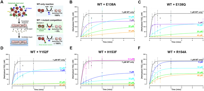
Eubacterial ribosomal large-subunit methyltransferase H (RlmH) methylates 23S ribosomal RNA pseudouridine 1915 (Ψ1915), which lies near the ribosomal decoding center. The smallest member of the SPOUT superfamily of methyltransferases, RlmH lacks the RNA recognition domain found in larger methyltransferases. The catalytic mechanism of RlmH enzyme is unknown. Here, we describe the structures of RlmH bound to S-adenosyl-methionine (SAM) and the methyltransferase inhibitor sinefungin. Our structural and biochemical studies reveal catalytically essential residues in the dimer-mediated asymmetrical active site. One monomer provides the SAM-binding site, whereas the conserved C-terminal tail of the second monomer provides residues essential for catalysis. Our findings elucidate the mechanism by which a small protein dimer assembles a functionally asymmetric architecture.
Conflict of interest statement
The authors declare that they have no competing interests.
Figures


Similar articles
-
Identification of pseudouridine methyltransferase in Escherichia coli.RNA. 2008 Oct;14(10):2223-33. doi: 10.1261/rna.1186608. Epub 2008 Aug 28. RNA. 2008. PMID: 18755836 Free PMC article.
-
YbeA is the m3Psi methyltransferase RlmH that targets nucleotide 1915 in 23S rRNA.RNA. 2008 Oct;14(10):2234-44. doi: 10.1261/rna.1198108. Epub 2008 Aug 28. RNA. 2008. PMID: 18755835 Free PMC article.
-
Specificity and kinetics of 23S rRNA modification enzymes RlmH and RluD.RNA. 2010 Nov;16(11):2075-84. doi: 10.1261/rna.2234310. Epub 2010 Sep 3. RNA. 2010. PMID: 20817755 Free PMC article.
-
Tied up in knots: Untangling substrate recognition by the SPOUT methyltransferases.J Biol Chem. 2022 Oct;298(10):102393. doi: 10.1016/j.jbc.2022.102393. Epub 2022 Aug 18. J Biol Chem. 2022. PMID: 35988649 Free PMC article. Review.
-
SAM (dependent) I AM: the S-adenosylmethionine-dependent methyltransferase fold.Curr Opin Struct Biol. 2002 Dec;12(6):783-93. doi: 10.1016/s0959-440x(02)00391-3. Curr Opin Struct Biol. 2002. PMID: 12504684 Review.
Cited by
-
Fragment-based discovery of a new class of inhibitors targeting mycobacterial tRNA modification.Nucleic Acids Res. 2020 Aug 20;48(14):8099-8112. doi: 10.1093/nar/gkaa539. Nucleic Acids Res. 2020. PMID: 32602532 Free PMC article.
-
Topologically knotted deubiquitinases exhibit unprecedented mechanostability to withstand the proteolysis by an AAA+ protease.Sci Rep. 2018 May 4;8(1):7076. doi: 10.1038/s41598-018-25470-0. Sci Rep. 2018. PMID: 29728659 Free PMC article.
-
RNA methylation in chloroplasts or mitochondria in plants.RNA Biol. 2021 Dec;18(12):2127-2135. doi: 10.1080/15476286.2021.1909321. Epub 2021 Apr 5. RNA Biol. 2021. PMID: 33779501 Free PMC article. Review.
-
SFP6 fluorescent probes for imaging SAM dynamics in living cells.Mikrochim Acta. 2025 Feb 21;192(3):180. doi: 10.1007/s00604-025-07039-7. Mikrochim Acta. 2025. PMID: 39982573
-
A Family Divided: Distinct Structural and Mechanistic Features of the SpoU-TrmD (SPOUT) Methyltransferase Superfamily.Biochemistry. 2019 Feb 5;58(5):336-345. doi: 10.1021/acs.biochem.8b01047. Epub 2018 Dec 3. Biochemistry. 2019. PMID: 30457841 Free PMC article.
References
Publication types
MeSH terms
Substances
Grants and funding
LinkOut - more resources
Full Text Sources
Other Literature Sources
Molecular Biology Databases
