. 2017 Apr 20;7(1):991.
doi: 10.1038/s41598-017-01116-5.

Transforming growth factor β plays an important role in enhancing wound healing by topical application of Povidone-iodine

Affiliations

Transforming growth factor β plays an important role in enhancing wound healing by topical application of Povidone-iodine

Li Wang et al. Sci Rep. .

Abstract

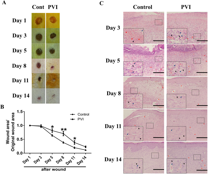
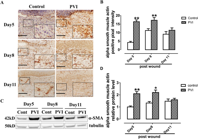
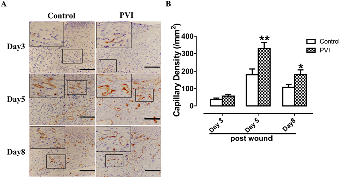
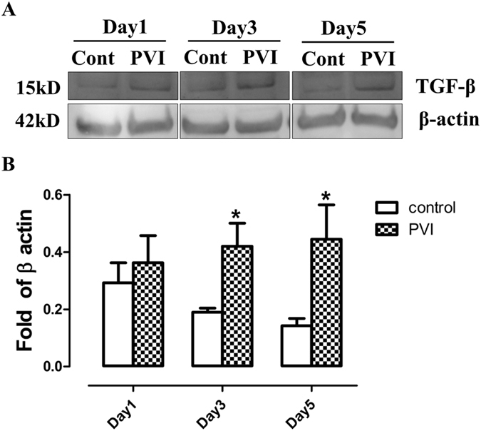
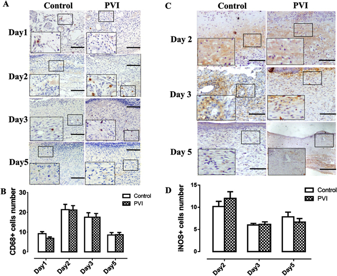
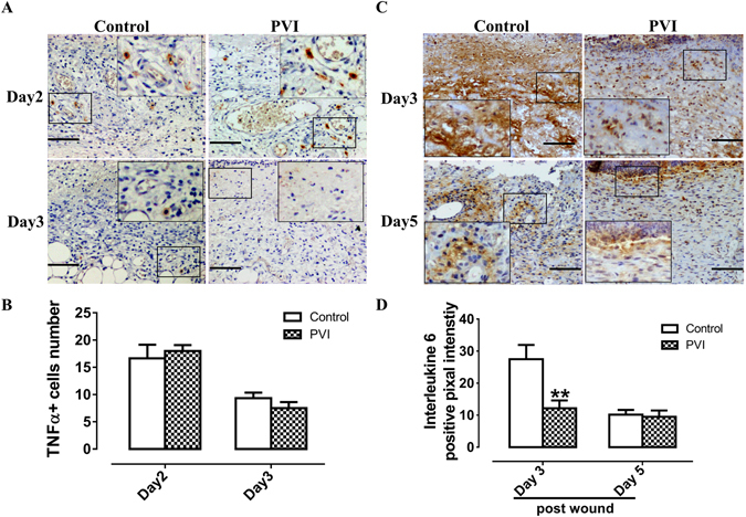
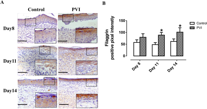
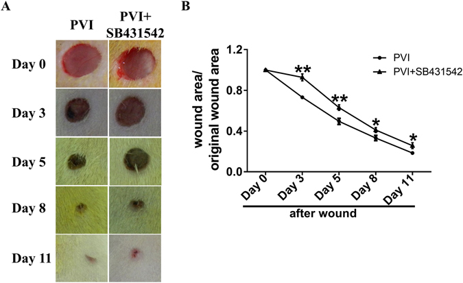
Povidone-iodine (PVI) is principally used as an antimicrobial agent. It has been found that 0.5% PVI can attenuate congestion, edema and pain induced by pressure sores. Thus this study aimed to assess the effects of 0.5% PVI on acute skin wounds. Four full-thickness excisional wounds were generated on the dorsal skin of male Sprague-Dawley rats with a 10-mm sterile punch. Two wounds were left untreated and the other two were dressed with gauze with 0.5% PVI for 1 hour per day for the first 5 days after injury. 10-mm full-thickness excisional wounds were also generated on the dorsal skin of rats treated with 10 mg/kg SB431542 and all wounds were treated with 0.5% PVI for 5 days. PVI treatment enhanced wound healing via promotion of expression of α SMA and TGF β, neovascularization and re-epithelialization. Interleukin 6 was reduced following PVI treatment. Inhibition of TGF β abolished the effect of PVI treatment on wound closure. These data show that topical application of 0.5% PVI could promote acute skin wound healing though increased expression of TGF β leading to enhanced formation of granulation tissue, even in the absence of obvious infection.


Conflict of interest statement

The authors declare that they have no competing interests.

Figures

Figure 1
Figure 1
Povidone-iodine (PVI) treatment enhanced cutaneous wound closure and granulation tissue formation and maturation. (A) Representative photographs of wounds from a control and PVI treated rat. (B) Wound contraction (%) at day 1 to 14 post-injury. (C) Representative micrographs of H&E stained wound sections. Red arrows indicate red blood cells. Blue arrows indicate inflammatory cells. Grey arrows indicate fibroblasts. Red asterisks indicate new blood vessels. Scale bar = 50 μm. *p < 0.05, **p < 0.01 vs control. Data are means ± S.D, n = 12.
Figure 2
Figure 2
Povidone-iodine (PVI) treatment promoted alpha smooth muscle actin (α-SMA) expression. (A) Representative immunohistochemical α-SMA stained wound sections from a control and PVI treated rat. Scale bar = 50 μm. (B) Semi-quantitative analysis of α-SMA expression. (C) Representative western blotting for α-SMA in wound areas. (D) Semi-quantitative analysis of α-SMA expression, n = 4. *p < 0.05, **p < 0.01 vs control. Data are means ± S.D, n = 24.
Figure 3
Figure 3
Povidone-iodine (PVI) treatment promoted wound neovascularization. (A) Representative immunohistochemical CD34 stained wound sections from a control and PVI treated rat. Scale bar = 50 μm. (B) Semi-quantitative analysis of capillary density (/mm2). Capillary density was assessed by counting the number of CD34 positive microvessels in high-power fields (40x). *p < 0.05, **p < 0.01 vs control. Data are means ± S.D, n = 24.
Figure 4
Figure 4
Povidone-iodine (PVI) treatment increased transforming growth factor β expression. (A) Representative western blotting for transforming growth factor (TGF) β in wound areas. (B) Semi-quantitative analysis of TGF-β expression. *p < 0.05 vs control. Data are means ± S.D, n = 4.
Figure 5
Figure 5
The effect of povidone-iodine (PVI) treatment on macrophages. (A) Representative immunohistochemical CD68 stained wound sections. Scale bar = 50 μm. (B) Quantitative analysis of CD68+ cells (/high power fields). (C) Representative immunohistochemical inducible nitric oxide synthase (iNOS) stained wound sections. Scale bar = 50 μm. (D) Quantitative analysis of iNOS + cells (/high power fields). Data are means ± S.D, n = 24.
Figure 6
Figure 6
The effect of povidone-iodine (PVI) treatment on tumor necrosis factor (TNF) α and interleukin (IL) -6 expression at the wound site. (A) Representative immunohistochemical TNFα stained wound sections. Scale bar = 50 μm. (B) Semi-quantitative analysis of TNFα + cells (/high power fields). (C) Representative immunohistochemical IL-6 stained wound sections. Scale bar = 50 μm. (D) Semi-quantitative analysis of IL-6 expression. **p < 0.01 vs control. Data are means ± S.D, n = 24.
Figure 7
Figure 7
Povidone-iodine (PVI) treatment enhanced filaggrin expression. (A) Representative immunohistochemical filaggrin stained wound sections. Scale bar = 50 μm. (B) Semi-quantitative analysis of filaggrin expression. *p < 0.05 vs control. Data are means ± S.D, n = 24.
Figure 8
Figure 8
Inhibition of transforming growth factor β prevented the promotion of skin wound healing by Povidone-iodine (PVI) treatment. (A) Representative photographs of wounds from rats treated with inhibitor of transform growth factor β (SB431542) or vehicle and PVI. (B) Wound contraction (%) at day 1 to 11 post-injury. *p < 0.05, **p < 0.01 vs control. Data are means ± S.D, n = 12.

Similar articles

Cited by

References

    1. Mimoz O, et al. Skin antisepsis with chlorhexidine-alcohol versus povidone iodine-alcohol, with and without skin scrubbing, for prevention of intravascular-catheter-related infection (CLEAN): an open-label, multicentre, randomised, controlled, two-by-two factorial trial. Lancet. 2015;386:2069–77. doi: 10.1016/S0140-6736(15)00244-5. - DOI - PubMed
    1. Noorani A, Rabey N, Walsh SR, Davies RJ. Systematic review and meta-analysis of preoperative antisepsis with chlorhexidine versus povidone-iodine in clean-contaminated surgery. Br J Surg. 2010;97:1614–20. doi: 10.1002/bjs.7214. - DOI - PubMed
    1. Shi CM, Nakao H, Yamazaki M, Tsuboi R, Ogawa H. Mixture of sugar and povidone-iodine stimulates healing of MRSA-infected skin ulcers on db/db mice. Arch Dermatol Res. 2007;299:449–56. doi: 10.1007/s00403-007-0776-3. - DOI - PubMed
    1. Shimamoto Y, Shimamoto H, Fujihata H, Nakamura H, Matsuura Y. Topical application of sugar and povidone-iodine in the management of decubitus ulcers in aged patients. Hiroshima J Med Sci. 1986;35:167–9. - PubMed
    1. Mitani O, et al. Enhanced wound healing by topical application of ointment containing a low concentration of povidone-iodine. J Wound Care. 2016;25:521–9. doi: 10.12968/jowc.2016.25.9.521. - DOI - PubMed

Publication types

LinkOut - more resources